Portable Handheld X-Ray Machine: Technology, Uses, Specifications & Buying Guide
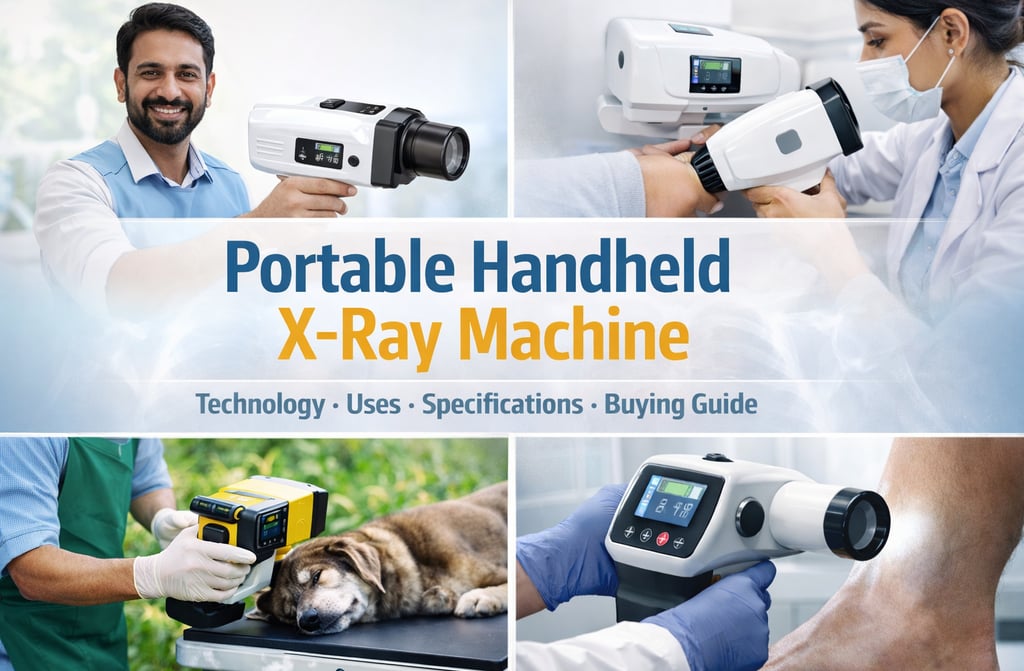
Portable handheld X-ray machines are compact diagnostic imaging devices used in hospitals, dental clinics, veterinary imaging and mobile healthcare units. Learn technology, specifications, applications and regulatory considerations.
HOT X-RAY PRODUCTS
3/10/20264 min read
Comprehensive Technical and Functional Profile of Portable Handheld X-Ray Machines
Portable handheld and ultraportable X-ray machines represent a significant technological shift in diagnostic imaging, moving from fixed room installations to flexible, point-of-care solutions. These devices are defined by the Atomic Energy Regulatory Board (AERB) as equipment intended to be moved between periods of use while being carried by one or two persons, with a strict weight limit not exceeding 12 kg. They are widely used in dental clinics, community-based chest X-ray (CXR) screening for Tuberculosis (TB), forensic operations, and emergency medical scenarios.
Core Functionality and Technical Specifications
Portable handheld units utilize high-frequency X-ray generators powered by high-capacity, rechargeable lithium-ion batteries. Unlike traditional units, they are often paired with digital sensors, photostimulable phosphor (PSP) plates, or highly sensitive LG Oxide detectors to produce diagnostic-quality images.
Key Technical Benchmarks
Technical specifications vary by clinical application, typically falling into two categories: dental intra-oral and general ultraportable (chest) systems.
Operating Potential (kVp) and Tube Current (mA):
Dental Handhelds (e.g., Nomad Pro): These typically operate at a constant potential of 60 kVp and a tube current of 2.5 mA.
Ultraportable Chest Systems: These offer a broader range, typically 40–100 kVp and 0.1–50 mAs. The World Health Organization (WHO) and International Atomic Energy Agency (IAEA) recommend a tube voltage capacity of at least 90 kV for chest imaging to ensure sufficient image contrast.
Focal Spot Size: Handheld units are engineered with smaller focal spots to maintain detail at lower power. For example, a standard handheld dental unit uses a 0.4 mm focal spot, whereas a traditional wall-mounted source often uses 0.7 mm.
Source-to-Skin Distance (SSD): Handheld units operate at shorter distances, typically 20 cm for dental intra-oral units compared to 30 cm for fixed units. For chest imaging, distances range from 1 m to 1.8 m.
Exposure Control: Units are equipped with preset timers that automatically terminate the X-ray beam after a specified duration to prevent unnecessary radiation exposure.
Performance and Image Quality Analysis
Research comparing handheld devices to fixed sources highlights specific advantages in resolution and dose efficiency.
Image Resolution (Line Pair Resolution)
A critical technical benchmark is the Line Pair (LP) resolution, which measures the ability of the system to visualize small details. Studies indicate that handheld devices often provide significantly higher mean LP resolution (6.05–6.55 lp/mm) compared to wall-mounted sources (5.58–6.31 lp/mm). This superior detail is primarily attributed to the smaller focal spot size.
Dosimetry and Dose Efficiency
Handheld units are designed to follow the ALARA (As Low As Reasonably Achievable) principle by optimizing exposure factors.
Patient Dose: In clinical simulations, the mean effective dose for a Full Mouth Examination (FMX) was found to be 36 µSv for handheld units, significantly lower than the 98 µSv recorded for wall-mounted devices. This represents a reduction of approximately 12% in total effective patient dose.
Operator Exposure: When used correctly, operator exposures from handheld units are often indistinguishable from ambient background levels (<2 µGy per study).
Design and Built-in Safety Features
Because the operator stands in close proximity to the X-ray source during handheld operation, the physical design of the machine includes specialized safety components.
Radiation Shielding
Backscatter Shielding: High-quality handheld units feature an integrated external lead-filled acrylic shield at the end of the collimator. This shield creates a "safe zone" for the operator by reflecting backscattered radiation away from them.
Internal Shielding: The X-ray tube housing is constructed to limit leakage radiation. For intra-oral units, leakage must not exceed 0.25 mGy in one hour at 1 meter. For general portable radiography, the limit is 1 mGy in one hour at 1 meter.
Safety Interlocks and Sensors
Proximity Sensors: Modern handheld devices incorporate proximity sensors that prevent the X-ray beam from switching "ON" unless the object to be scanned is at the intended distance.
Fail-Safe Mechanisms: The machine is designed to automatically de-energize in the event of a malfunction. Exposure can also be manually terminated at any moment via the trigger.
Key/Password Control: To prevent unauthorized use, operation is often key-controlled or password-protected.
Technical Pros and Cons of Handheld Systems
Pros
Portability and Mobility: These machines allow for diagnostics in field conditions, forensics, and humanitarian missions where traditional infrastructure (like stable power) is absent.
Operator Satisfaction: Clinical studies report that 97% of operators find handheld devices easier to use compared to 50% for wall-mounted sources.
Technique Efficiency: Handheld units reduce the need for patient movement, which is particularly beneficial for pediatric, geriatric, or trauma patients.
Cons
Radiation Geometry: The operator stands near the source, which requires strict adherence to shielding protocols and distance rules.
Physical Fatigue: Repeated one-handed use of a device (even under 12 kg) can lead to operator fatigue during high-volume screening.
Higher Regulatory Risk: Under India’s Medical Device Rules 2017, these machines are classified as Class C (Moderate to High Risk) medical devices, requiring mandatory CDSCO registration and AERB Type Approval due to the potential risks of misdiagnosis or inferior quality in non-regulated units.
Quality Assurance (QA) Benchmarks
To ensure consistent functional performance, handheld X-ray machines must meet specific AERB-mandated QA tolerances.
Mandatory QA Tests and Tolerances
Accelerating Potential (kVp): The delivered kVp must be within ± 5 kV of the set value.
Timer Accuracy: The exposure time error must not exceed ± 10%.
Output Consistency: The coefficient of variation (CoV) for radiation output must be < 0.05.
Linearity of Radiation Output: The coefficient of linearity (CoL) for mA and timer loading stations must be < 0.1.
Minimum Total Filtration: To cut off low-energy components that do not contribute to the image, the total filtration must be:
1.5 mm Al for units operating at ≤ 70 kV.
2.0 mm Al for units between 70–100 kV.
2.5 mm Al for units > 100 kV.
Machine Components and Accessories
A complete ultraportable handheld system typically includes the following major components:
X-Ray Generator: Features an inbuilt battery and wireless remote control accessories.
Digital Detector: Often a LG Oxide Detector (e.g., 14” x 17”) with extra batteries and a dedicated charger.
Portable Workstation: A laptop or PC-console with pre-installed CAD (Computer-Aided Detection) software, often powered by AI for automated image interpretation.
Support Hardware: Tripod stands for both the generator and detector to ensure stability during exposures.
Protection Gear: Mandatory lead aprons (minimum 0.25 mm lead equivalence) and TLD badges for personnel monitoring.
Transportation Case: A hard case with a number-coded lock for secure transport and storage.
Conclusion
Portable handheld X-ray machines are "hot" diagnostic products because they combine high-resolution imaging with extreme mobility. However, their technical superiority is contingent upon strict adherence to AERB QA benchmarks, the maintenance of internal and external shielding, and rigorous Class C regulatory compliance.
Portable handheld X-ray machines have become an important tool in modern diagnostic imaging. Their compact design, digital compatibility and mobility allow healthcare professionals to perform radiographic examinations in situations where traditional radiology equipment may not be practical.
As imaging technology continues to evolve, portable radiography devices are expected to contribute significantly to expanding access to medical diagnostics.
About XRAYNEWS.NET
XRAYNEWS.NET provides updates on medical and industrial X-ray technologies, radiography insights, regulatory awareness, and imaging industry developments.
Regulatory Resources
© 2025. All rights reserved.
Video Resources
Popular Topics
Quick Links
Stay Updated with X-Ray Industry News
Subscribe to receive updates on X-ray technology developments, radiography insights, and industry news.