Low-Dose Imaging in Radiology: Advancing Patient Safety & Diagnostic Excellence
Explore the latest developments in low-dose imaging technology focused on patient safety and radiological excellence. Learn how ALARA principles, advanced X-ray systems, and AERB & CDSCO compliance are reducing radiation exposure while maintaining high diagnostic accuracy.
4/26/20265 min read
Low-Dose Imaging: New Developments in Patient Safety and Radiological Excellence
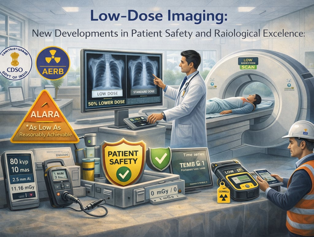
In the evolving field of medical diagnostics, the use of X-rays has proven immensely beneficial for the diagnosis and treatment of various ailments. However, because ionizing radiation carries inherent health risks, it is a legal and ethical mandate that exposures be kept As Low As Reasonably Achievable (ALARA). Modern developments in low-dose imaging are centered on this principle, ensuring that the diagnostic task is matched to the imaging modality so that patient benefit outweighs any risk.
The radiological landscape in India is currently defined by a synergy between high-tech innovations and a rigorous regulatory framework led by the Atomic Energy Regulatory Board (AERB) and the Central Drugs Standard Control Organization (CDSCO). Together, they ensure that patient safety is "built-in" by design and maintained through religious operational protocols.
1. The Technological Shift to Low-Dose Hardware
A significant development in patient safety is the transition from conventional wall-mounted X-ray sources to advanced handheld and high-frequency units.
Handheld vs. Wall-Mounted Systems: A Dosimetry Breakthrough
Independent research comparing handheld devices, such as the Nomad Pro, with traditional wall-mounted units has revealed significant advantages in dose reduction. Clinical simulations of a Full Mouth Examination (FMX) demonstrated that the mean patient dose was significantly less for handheld devices (36µSv) compared to wall-mounted devices (98µSv). This represents a reduction in total effective patient dose of approximately 12%.
Optimization of Exposure Factors: This dose reduction is primarily attributed to the optimization of exposure factors such as kilovoltage (kVp), milliamperage (mA), and exposure time.
High-Frequency Generators: Unlike conventional units that may produce pulsating waveforms, modern handheld units utilize high-frequency constant potential generators. This ensures the tube operates at the peak voltage throughout the exposure, eliminating "soft" low-energy X-rays that increase patient dose without aiding image formation.
Focal Spot Size and Image Resolution
Patient safety is also enhanced by the ability to achieve superior image quality at lower doses. Handheld devices often feature a smaller focal spot size (0.4 mm) compared to fixed units (0.7 mm). A smaller focal spot results in significantly higher Line Pair (LP) resolution, allowing clinicians to resolve finer anatomical details with less radiation.
2. Ultra-Portable (UP) Systems: Decentralizing Safe Care
One of the "hottest" trends in low-dose imaging is the rise of Ultra-portable (UP) X-ray systems, which fit within a suitcase or backpack and can be moved regularly to areas of need.
Portability Standards: The AERB defines portable equipment as units intended to be moved between locations by one or two persons, with a weight not exceeding 12 kg.
Infrastructure Independence: These systems are composed of a low-weight, battery-powered X-ray tube and a dose-efficient digital detector, making them ideal for settings where conventional infrastructure (like a stable power supply) is unavailable.
Field Safety: In community settings, where lead-lined rooms are absent, safety is maintained by ensuring a cordoned-off area so that the total dose to a person at the boundary (30 meters from the generator) does not exceed 5 µSv per scan.
3. AI and CAD: Standardizing Low-Dose Diagnostics
The integration of Artificial Intelligence (AI) and Computer-Aided Detection (CAD) software with UP systems has transformed how India manages public health crises like Tuberculosis (TB).
Automated Triage: CAD tools provide automated and standardized image interpretation, which is vital in regions where expert human readers are scarce.
Standardized Quality: By using AI to highlight the likelihood of abnormalities in a Chest X-ray (CXR), these systems facilitate rapid triage for molecular tests while ensuring that exposure parameters are consistently optimized.
Cloud-Based PACS: Modern implementations utilize cloud-based Picture Archiving and Communication Systems (PACS) to store scans for over three years, ensuring diagnostic data is never lost or unintentionally altered.
4. Regulatory Framework: Class C Classification and e-LORA
A landmark shift occurred on January 1, 2021, when the CDSCO mandated that all X-ray machines in India be registered as Class C (Moderate to High Risk) medical devices.
Preventing Misdiagnosis: This regulation was implemented to curb a "big spurt" in misbranded and inferior-quality products that lead to clinical misdiagnosis and inaccurate reporting.
The e-LORA Gateway: All regulatory transactions—from procurement permissions to operational licenses—are managed via the e-Licensing of Radiation Applications (e-LORA) digital portal.
Mandatory Licensing: No diagnostic X-ray unit can be legally operated for patient diagnosis until a Licence for Operation is obtained from the AERB.
5. Quality Assurance (QA): The Backbone of Patient Protection
Safety is not just about having high-spec hardware; it requires continuous verification that the equipment is performing within mandatory tolerances.
Critical QA Parameters for Low-Dose Imaging
Tube Potential (kVp) Accuracy: Must be within ± 5 kV of the set value to ensure consistent beam penetration.
Exposure Timer Accuracy: Inaccurate timers can lead to poor contrast or motion artifacts; the acceptable error is within ± 10%.
Minimum Total Filtration: To absorb useless, low-energy radiation, the total filtration must be at least 2.5 mm Al equivalent for constant potential equipment.
Central Beam Alignment: If the X-ray beam is not perpendicular to the image receptor (aligned within < 1.5°), the image may be distorted, requiring a patient retake and doubling their dose.
6. Operational Safety and the TDS Principle
The human element of safety remains the most effective tool in dose reduction. All facilities must foster a "Safety Culture" based on the Time-Distance-Shielding (TDS) principle.
Time: Operators should minimize the total "beam-on" time.
Distance: For mobile and portable units, the operator is mandated to maintain a distance of at least 2 meters from the source during exposure.
Shielding: Physical barriers are essential for protection against scattered radiation.
Lead Aprons: Must have a minimum of 0.25 mm lead equivalence.
Mobile Protective Barriers (MPB): Require a minimum of 1.5 mm lead equivalence.
Structural Shielding: All doors to X-ray rooms must have a lead lining of 1.7 mm.
7. Roles and Responsibilities in the Safety Chain
The AERB distributes accountability among several key stakeholders to ensure patient and public safety.
The Employer: Acts as the custodian of the equipment and is responsible for procuring only AERB Type Approved models from authorized suppliers.
The Radiological Safety Officer (RSO): A qualified professional approved by the Competent Authority to implement radiation surveillance and advise on modifying working conditions for pregnant radiation workers.
The Licensee: Responsible for maintaining dose records and arranging periodic Quality Assurance tests every two years.
The Radiation Worker: Personnel mandated to wear Thermoluminescent Dosimeter (TLD) badges below the lead apron at chest level to track their annual effective dose limit of 20 mSv.
Conclusion: Achieving Diagnostic Excellence
Developments in low-dose imaging—from high-frequency generators and ultra-portable UP systems to AI-powered CAD tools—are revolutionizing patient safety in India. By focusing on the ALARA principle and adhering to the rigorous CDSCO and AERB regulatory frameworks, medical facilities can bridge the diagnostic gap while upholding the highest standards of care.
A good diagnostic procedure is one that provides the optimum quality diagnostic information at the lowest possible radiation risk. Success in this modern era depends on religious adherence to biennial Quality Assurance, meticulous e-LORA licensing, and a commitment to the Time-Distance-Shielding principle. Failure to comply with these guidelines is a punishable offense under the Atomic Energy Act, 1962, underscoring that in the field of radiology, safety is the primary mandate for public trust and clinical excellence.
About XRAYNEWS.NET
XRAYNEWS.NET provides updates on medical and industrial X-ray technologies, radiography insights, regulatory awareness, and imaging industry developments.
Regulatory Resources
© 2025. All rights reserved.
Video Resources
Popular Topics
Quick Links
Stay Updated with X-Ray Industry News
Subscribe to receive updates on X-ray technology developments, radiography insights, and industry news.