Wireless DR Panels in Radiology: Latest Innovations, Benefits & Use Cases
Explore the latest advancements in wireless DR panels in modern radiology. Learn how portable digital detectors improve imaging speed, workflow efficiency, and patient care while ensuring AERB compliance and high-quality diagnostics in hospitals and remote settings.
4/28/20264 min read

Wireless DR Panels: Latest Improvements and Use Cases in Modern Radiology
The landscape of diagnostic imaging is currently undergoing a rapid digital transformation, shifting away from traditional film-based and Computed Radiography (CR) systems toward Direct Digital Radiography (DR). At the heart of this evolution is the Imaging Device, defined by the Atomic Energy Regulatory Board (AERB) as a detector unit or array of detectors that receives X-rays and responds by producing an electrical or light signal.
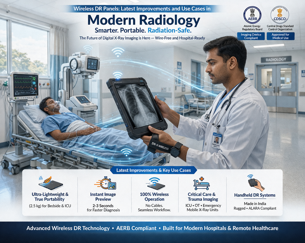
The introduction of Wireless DR Panels—particularly those integrated into ultra-portable and handheld systems—has revolutionized medical diagnostics by enabling high-quality imaging in diverse settings, from metropolitan hospitals to remote community health camps. This guide explores the technological advancements, regulatory requirements, and high-impact use cases of wireless DR technology in the context of the Indian radiological ecosystem.
1. The Technology Behind Wireless DR Panels
Modern wireless DR panels represent the pinnacle of "built-in safety" and functional excellence. Unlike older CR systems that utilize Photostimulable Phosphor (PSP) plates requiring a separate scanning step at resolutions like 300 DPI, wireless DR panels provide near-instantaneous visualization.
A. Scintillator and Detector Tech
State-of-the-art panels, such as the LG Oxide Detector (commonly sized at 14” x 17”), use highly sensitive materials to convert X-ray energy into digital data. These detectors are designed to be dose-efficient, meaning they can produce high-contrast images with significantly lower radiation exposure compared to analog film, adhering to the international ALARA (As Low As Reasonably Achievable) principle.
B. Portability and Power
Latest improvements focus on miniaturization and power management. Modern detectors are battery-powered and often supplied with extra detector batteries and specialized charging stations to ensure uninterrupted field operation. AERB defines Portable X-ray equipment as units weighing not more than 12 kg, a benchmark made possible by these lightweight digital panels.
2. Latest Improvements in Digital Imaging
Recent advancements in wireless DR technology have focused on enhancing spatial resolution and operational efficiency.
Higher Resolution (Line Pair/mm): Research comparing digital handheld devices with traditional wall-mounted sources indicates that modern digital systems achieve significantly higher mean Line Pair (LP) resolution. While traditional fixed units may resolve around 5.58–6.31 lp/mm, high-spec digital systems paired with small focal spot generators (0.4 mm) can achieve 6.05–6.55 lp/mm, allowing for the visualization of minute anatomical details.
Detector Characteristics: Modern panels feature improved Detective Quantum Efficiency (DQE), which enhances the signal-to-noise ratio. This allows for clear images even when manual exposure adjustments (kVp and mAs) are required in field settings where Automatic Exposure Control (AEC) is absent.
Wireless Connectivity and Speed: Advanced panels utilize high-speed Wi-Fi protocols to transmit large DICOM files to a portable workstation or laptop in seconds. This allows for on-the-spot quality checks and rapid diagnosis.
3. Strategic Use Cases: From Bedside to the Community
The mobility of wireless DR panels has expanded the reach of radiology far beyond the lead-lined walls of traditional departments.
A. Community-Based TB Screening
The "main application" of ultra-portable wireless systems today is Chest X-ray (CXR) screening for pulmonary Tuberculosis (TB).
AI-CAD Integration: These panels are frequently paired with Artificial Intelligence (AI) and Computer-Aided Detection (CAD) software, which provides automated, standardized interpretation.
Triage Efficiency: AI highlights abnormalities suggestive of TB, allowing non-expert staff to triage patients for molecular NAAT testing, which is vital in regions where expert human readers are scarce.
B. Emergency and Field Operations
Wireless DR technology is the "ideal tool" for:
Bedside Imaging: Providing diagnostics for immobilized patients in emergency rooms or temporary field hospitals.
Forensic and Combat Ops: Used by military and disaster response teams where traditional fixed sources are unavailable.
Security Inspections: Portable X-ray Scanners (PXS) are used for imaging suspicious items in the field, with remote operation capabilities from at least 30 meters away.
4. Regulatory Framework and E-E-A-T in India
Operating wireless DR panels in India requires a deep commitment to Expertise, Authoritativeness, and Trustworthiness (E-E-A-T), as enforced by the AERB and CDSCO.
CDSCO Classification: Since January 1, 2021, all X-ray Generating Equipment (XGE) and associated digital detectors are classified as Class C (Moderate to High Risk) medical devices. Registration is mandatory to prevent the use of misbranded or inferior products that could lead to misdiagnosis.
The e-LORA Portal: All regulatory transactions—from obtaining a Procurement Letter to receiving a Licence for Operation—must be managed through the e-Licensing of Radiation Applications (e-LORA) digital portal.
Institutional Roles: Every facility must designate an AERB-approved Radiological Safety Officer (RSO) to oversee safety and maintain dose records.
5. Quality Assurance (QA) and Maintenance
To maintain diagnostic precision, wireless DR panels must undergo rigorous periodic testing.
A. Periodicity and Calibration
Biennial QA: The end-user must ensure that periodic Quality Assurance is carried out by authorized agencies once every two years.
Software Calibration: Digital detectors must be calibrated periodically as per the manufacturer’s recommendations to ensure that functional performance remains similar to baseline values.
B. Technical Benchmarks
QA tests verify parameters such as:
kVp Accuracy: Within ± 5 kV of the set value.
Exposure Timer Accuracy: Percentage error not to exceed ± 10%.
Uniformity: Ensuring the panel produces a uniform image without artifacts.
6. Digital Workflow: PACS and Cloud Imaging
The final component of modern digital radiology is the management of the data captured by the DR panel.
PACS (Picture Archiving and Communication System): Modern systems use PACS software to store, retrieve, and distribute images. AERB mandates that PACS must ensure patient information is not lost or unintentionally altered.
Cloud-Based Storage: Recent implementations, particularly in TB screening programs, utilize cloud-based systems to store scans for over 3 years, facilitating telemedicine and remote second opinions.
Interconnectivity: Systems must exchange information in accordance with national and international standards (such as DICOM) to ensure data integrity across computer networks.
7. Essential Radiation Safety Protocols
While technology has made imaging safer, the human operator must still adhere to the Time-Distance-Shielding (TDS) principle.
Safe Distance: For portable and mobile operations, the operator is mandated to maintain a distance of at least 2 meters from the source during activation.
Cordoning Areas: In community field settings where lead-lined rooms are unavailable, an area must be cordoned off so the total dose to a person at 30 meters does not exceed 5 µSv per scan.
Personal Protection: Radiographers must use TLD (Thermoluminescent Dosimeter) badges (worn below the lead apron) to track their annual whole-body dose, which is limited to 20 mSv averaged over five years.
Conclusion
Wireless DR panels are more than just a hardware upgrade; they are the foundation of a safer, more efficient, and more accessible diagnostic future. By integrating high-resolution digital detection with battery-powered mobility and AI-driven triage, these systems are successfully bridging the diagnostic gap in India.
However, the success of this technology depends on religious adherence to regulatory standards. By maintaining biennial Quality Assurance, ensuring meticulous e-LORA licensing, and upholding the ALARA principle, medical facilities can guarantee that diagnostic information is obtained at the lowest possible radiation risk to the patient. Compliance with the Atomic Energy Act, 1962 is not merely a legal requirement; it is a commitment to public health and clinical excellence.
About XRAYNNEWS.NET
XRAYNEWS.NET provides updates on medical and industrial X-ray technologies, radiography insights, regulatory awareness, and imaging industry developments.
Regulatory Resources
© 2025. All rights reserved.
Video Resources
Popular Topics
Quick Links
Stay Updated with X-Ray Industry News
Subscribe to receive updates on X-ray technology developments, radiography insights, and industry news.