AI-Powered Radiology in India: How Artificial Intelligence is Transforming X-Ray Diagnostics & TB Screening
Explore how AI-powered radiology and CAD software are transforming X-ray diagnostics in India with faster detection, improved accuracy, and AERB-compliant safety in TB screening programs.
INDUSTRY INSIGHTS & TECHNOLOGY
3/18/20265 min read
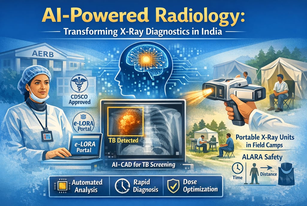
AI-Powered Radiology: How Artificial Intelligence is Transforming X-Ray Diagnostics in India
The mission of the Atomic Energy Regulatory Board (AERB) is to ensure that the use of ionizing radiation in India does not cause undue risk to the health of people and the environment. In recent years, this mission has intersected with a technological revolution: the integration of Artificial Intelligence (AI) and Computer-Aided Detection (CAD) software with modern X-ray systems. This transformation is particularly visible in the deployment of ultra-portable (UP) and handheld X-ray devices for community-based screening. By automating image interpretation and optimizing dose efficiency, AI is bridging the gap between advanced diagnostics and resource-limited settings, fundamentally changing how India manages public health crises like Tuberculosis (TB).
1. The Regulatory Landscape: From X-Rays to "Drugs"
In India, the regulatory framework for radiation safety is established by the Atomic Energy (Radiation Protection) Rules, 2004, promulgated under the Atomic Energy Act, 1962. A pivotal shift occurred on January 1, 2021, when the Central Drugs Standard Control Organization (CDSCO) classified all X-ray generating equipment as Class C (Moderate to High Risk) medical devices.
Key Regulatory Mandates:
Mandatory Registration: All manufacturers and importers must register X-ray machines with the CDSCO to prevent the proliferation of misbranded or inferior-quality products that lead to misdiagnosis.
AERB Type Approval: Prior to commercial production or marketing, every model must obtain a Type Approval Certificate, verifying it meets national safety standards.
e-LORA Portal: All regulatory transactions—from procurement permissions to operational licenses—are managed via the e-Licensing of Radiation Applications (e-LORA) portal.
2. The Rise of AI-Powered Computer-Aided Detection (CAD)
Ultra-portable X-ray systems have proven uniquely useful when paired with AI-powered CAD software. This combination allows for automated and standardized image interpretation without the constant need for expert human readers, who are often scarce in low- and middle-income countries (LMIC).
A. Transforming TB Screening
Currently, the primary application for UP systems with CAD tools is Chest X-ray (CXR) screening for pulmonary TB. TB has the highest annual mortality of all infectious diseases despite being treatable; early diagnosis is critical and achieved through systematic, large-scale screening to triage patients for molecular tests.
Likelihood Highlighting: In field settings, AI highlights the likelihood of a CXR being suggestive of TB or other chest abnormalities, allowing non-radiographer staff to facilitate triage.
Standardized Interpretation: CAD tools provide a level of consistency that is vital for high-throughput screening programs, such as those implemented in districts supported by the William J. Clinton Foundation (WJCF) and the Ministry of Health and Family Welfare.
3. Hardware Evolution: Handheld and Ultra-Portable Systems
Modern X-ray technology is moving away from traditional fixed sources toward compact, battery-powered units that fit within a suitcase or backpack.
Handheld vs. Wall-Mounted Performance: Research comparing handheld devices (like the Nomad Pro) to traditional wall-mounted sources reveals significant technical advantages:
Focal Spot Size: Handheld units often feature a smaller focal spot (0.4 mm) compared to fixed units (0.7 mm).
Image Detail: A smaller focal spot results in significantly higher mean Line Pair (LP) resolution, allowing for finer anatomical detail.
Generator Efficiency: Handheld units utilize high-frequency constant potential generators (e.g., 60 kVp), which eliminate low-energy "soft" X-rays that increase patient dose without aiding the image.
4. Radiation Safety and the ALARA Principle
Every radiation installation in India must adhere to the ALARA (As Low As Reasonably Achievable) principle. This ensures that exposures are kept as low as practicable, taking economic and social factors into account.
A. Occupational Dose Limits
The AERB sets strict annual effective dose limits to protect radiation workers:
Occupational Workers: 20 mSv per year averaged over five years.
Public Exposure: 1 mSv in a year.
Pregnant Workers: Once pregnancy is notified, the embryo/foetus should not receive more than 1 mSv for the remainder of the pregnancy.
B. Protective Protocols
AI systems help optimize safety by reducing the need for "retakes" through precise exposure calibration. However, physical protection remains mandatory:
Lead Aprons: Operators must wear aprons with a minimum 0.25 mm lead equivalence.
TLD Badges: Personnel monitoring via Thermoluminescent Dosimeter (TLD) badges is required for all radiation workers. These badges must be worn below the lead apron at chest level.
Time-Distance-Shielding (TDS): Increasing the distance from the source is the most effective way to reduce scatter radiation. For mobile units, the exposure cable length must be at least 2 meters.
5. Field Safety and Public Protection
Deploying AI-powered X-ray systems in the community (Active Case Finding camps) presents unique challenges because conventional lead-lined rooms are unavailable.
Safety Guidelines for Field Operations:
Cordoning Off Area: For portable scanners, an area must be cordoned off so that the total dose to a person at the boundary (30 meters from the generator) does not exceed 5 µSv per scan.
Scattered Radiation: The highest scatter dose is observed in the direction back towards the X-ray tube (180°/backscatter). Operators should maintain a safe distance—calculations show a workload of 50 exams/day requires a distance of 4.5 meters if no shielding is used.
Environmental Protection: Storage facilities for these devices should be sanitary, free from dust, water leakages, and direct sunlight.
6. Institutional Roles and Responsibilities
The Employer (e.g., a District Hospital or Diagnostic Center) holds ultimate responsibility for ensuring radiation safety and acting as the custodian of the equipment.
A. The Radiological Safety Officer (RSO)
Every facility must designate an AERB-approved RSO.
Qualifications: For high-risk facilities like CT or Interventional Radiology, the RSO must be a radiologist or a technologist with three years of experience.
Duties: The RSO maintains Quality Assurance (QA) records, advises on safety measures, and conducts radiation protection surveys.
B. Quality Assurance (QA) Benchmarks
The end-user is responsible for ensuring that periodic QA is carried out by authorized agencies once every two years.
kVp Accuracy: Must be within ± 5 kV of the set value.
Timer Accuracy: Percentage error must not exceed ± 10%.
Leakage Radiation: For radiography units, leakage through the housing must be < 1 mGy in one hour at 1 meter from the focus.
7. Procurement and Licensing via e-LORA
A facility cannot legally operate an X-ray unit for patient diagnosis until it obtains a Licence for Operation from the Competent Authority.
The Step-by-Step Pathway:
Institute Registration: The facility registers on e-LORA using valid credentials and identifies the Employer and Licensee.
Procurement Letter: The Employer applies for a Procurement Letter by selecting an AERB Type Approved model and an authorized supplier (e.g., Wipro GE, Siemens, or Fujifilm).
Installation and QA: After delivery, an authorized engineer installs the device and generates an installation report. A third-party agency then conducts a QA check.
Operational License: The installation and QA reports are uploaded to e-LORA. Approval typically takes 30 days, and the license must be carried to all field camps.
8. Challenges and Future Outlook
While AI is transforming diagnostics, the study of UP systems highlights a critical need for updated radiation safety guidelines specifically for field use. Traditional standards designed for lead-lined hospital rooms can restrict the potential of mobile units.
Future Priorities:
Training: Ensuring all staff working with AI-CAD systems, including non-radiographers, receive formal training on optimization and collimation to reduce unnecessary dose.
AI for Optimization: Developing AI that can automatically adjust exposure factors (mAs) based on patient size, as most current UP devices lack Automatic Exposure Control (AEC).
PACS Integration: Ensuring digital systems exchange information according to national and international standards to prevent the loss of patient data.
Conclusion
AI-powered radiology is not merely a technical upgrade; it is a vital public health tool for India. By combining the mobility of ultra-portable X-ray systems with the standardized interpretation of CAD tools, India can bring high-quality diagnostic services to the most remote populations. However, this expansion must be grounded in the authoritative regulatory framework provided by the AERB and CDSCO. Through biennial Quality Assurance, strict e-LORA licensing, and a religious commitment to the ALARA principle, India can leverage AI to eliminate diseases like TB while ensuring the absolute safety of patients, staff, and the public. Compliance with national safety codes is the hallmark of a facility that prioritizes diagnostic excellence and human health.
About XRAYNNEWS.NET
XRAYNEWS.NET provides updates on medical and industrial X-ray technologies, radiography insights, regulatory awareness, and imaging industry developments.
Regulatory Resources
© 2025. All rights reserved.
Video Resources
Popular Topics
Quick Links
Stay Updated with X-Ray Industry News
Subscribe to receive updates on X-ray technology developments, radiography insights, and industry news.