How X-Ray Imaging Supports TB Detection and Public Health Programs
Discover how X-ray imaging plays a vital role in tuberculosis (TB) detection and public health programs. Learn about chest X-ray screening, early diagnosis, mobile radiology units, and safety standards ensuring effective TB control and patient protection.
MEDICAL X-RAY UPDATES
4/27/20265 min read
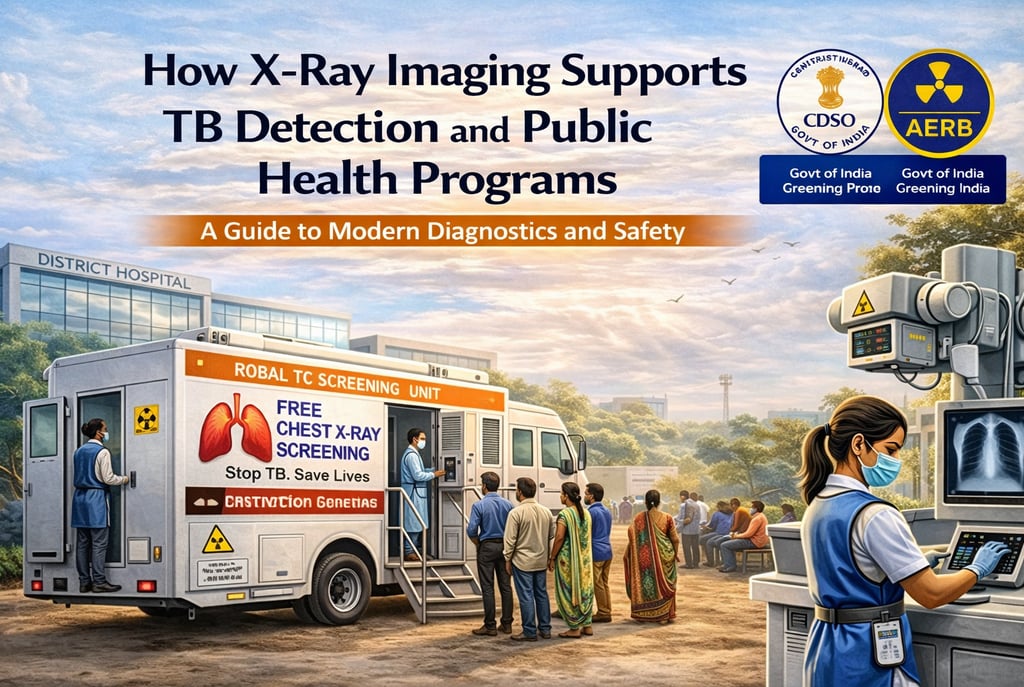
How X-Ray Imaging Supports TB Detection and Public Health Programs: A Guide to Modern Diagnostics and Safety
Tuberculosis (TB) remains one of the world's deadliest infectious diseases, despite being treatable. In the effort to eliminate TB, the medical use of X-rays for diagnosis has proven immensely beneficial to society. Systematic and large-scale Chest X-ray (CXR) screening is now a critical tool for identifying presumptive cases in high-risk subpopulations and triaging them for molecular tests. In India, these public health efforts are governed by the Atomic Energy Regulatory Board (AERB) and the Central Drugs Standard Control Organization (CDSCO) to ensure that the benefits of radiation diagnostics outweigh the inherent biological risks.
1. The Shift to Ultra-Portable (UP) X-Ray Systems
Traditionally, TB screening was restricted to hospital environments with fixed X-ray machines. Recent advancements have led to the development of Ultra-Portable (UP) X-ray devices, which are designed to fit within a suitcase or backpack and can be moved regularly to areas of need.
System Components: These units typically consist of a low-weight, battery-powered X-ray tube and a highly sensitive digital detector.
Infrastructure Independence: A major advantage of UP systems is their ability to function in settings where conventional infrastructure, such as a stable power supply or dedicated lead-lined rooms, is unavailable.
Technological Benchmarks: Research on similar handheld technology, like the Nomad Pro, shows that modern portable units can utilize high-frequency constant potential generators (e.g., 60 kVp) to optimize radiation quality and reduce unnecessary "soft" radiation.
Image Clarity: Portable units often feature a smaller focal spot (0.4 mm) compared to fixed units (0.7 mm), which results in significantly higher Line Pair (LP) resolution. This allows for the visualization of minute anatomical details critical for detecting early TB lesions.
2. AI-Powered Triage and Public Health Integration
The integration of Artificial Intelligence (AI) and Computer-Aided Detection (CAD) software has revolutionized TB screening, particularly in low- and middle-income countries (LMIC) where expert human readers are scarce.
Automated Interpretation: CAD tools provide automated and standardized image interpretation, highlighting the likelihood of a CXR being suggestive of TB or other chest abnormalities.
Reducing Stigma: Public health programs in India suggest setting up TB screening camps as "General Health Camps". Offering additional services like BMI checks and blood pressure monitoring helps reduce the stigma associated with TB in the community.
Active Case Finding (ACF): The Ministry of Health and Family Welfare utilizes these units for ACF surveys, reaching vulnerable groups through direct door-to-door visits and congregation spots.
Telemedicine Support: AI outputs are often supplemented by telemedicine, allowing remote experts to confirm findings and initiate treatment requests via systems like Ni-kshay.
3. Radiation Safety in Community Settings
When X-ray screening moves from lead-lined rooms to open community settings, strict adherence to radiation safety principles—Time, Distance, and Shielding (TDS)—is mandatory.
A. Scattered and Leakage Radiation
Even though the primary beam is focused on the patient, radiographers and the public are exposed to scattered radiation from the patient and leakage radiation from the tube housing.
Backscatter: The highest scatter dose is observed in the direction back towards the X-ray tube (180°).
Safe Distances: Operators are mandated to maintain a distance of at least 2 meters from the equipment during activation.
Cordoning Off Areas: For field scanners, a boundary should be cordoned off so that the dose to a person at 30 meters does not exceed 5 µSv per scan.
B. Personnel Protection
Lead Aprons: All radiation workers must wear protective aprons with a minimum of 0.25 mm lead equivalence.
Personnel Monitoring: Workers are required to wear Thermoluminescent Dosimeter (TLD) badges below the lead apron at chest level. These badges are exchanged quarterly for dose reporting to ensure exposures remain below the annual limit of 20 mSv.
Pregnant Workers: Once a female worker declares her pregnancy, her working conditions must be modified to ensure the embryo/foetus dose does not exceed 1 mSv for the remainder of the term.
4. Regulatory Compliance and e-LORA Portal
Operating X-ray equipment in India requires a rigorous consenting process handled via the e-Licensing of Radiation Applications (e-LORA) portal.
CDSCO Classification: Since January 1, 2021, all X-ray machines are classified as Class C (Moderate to High Risk) medical devices, making mandatory registration a legal prerequisite to prevent the use of inferior-quality products.
Procurement Permission: The Employer (the person with overall responsibility for the facility) must obtain a Procurement Letter via e-LORA before purchasing any X-ray unit.
Operational License: No diagnostic X-ray equipment shall be operated for patient diagnosis until a Licence for Operation is obtained from the Competent Authority.
Type Approval: Every model must obtain a Type Approval Certificate based on a demonstration of satisfactory performance by a prototype. This approval becomes invalid if design specifications are altered.
5. Operational Guidance for TB Programs
The successful deployment of UP systems for TB detection depends on a coordinated effort between various stakeholders.
Key Stakeholder Roles
State TB Officer (STO): Responsible for site identification and monitoring outcomes at the state level.
District TB Officer (DTO): Acts as the custodian of the equipment, responsible for registration and receiving HHXray machines.
Radiological Safety Officer (RSO): A qualified professional (e.g., a radiologist or technologist with three years of field experience) approved by AERB to oversee radiation protection.
Radiographer: Responsible for the overall handling and operation of the HHXray device in the field.
Maintenance and Quality Assurance (QA)
Biennial Testing: The end user must ensure that periodic Quality Assurance tests are carried out by authorized agencies once every two years.
QA Parameters: Essential checks include operating potential (kVp) accuracy within ± 5 kV and timer accuracy within ± 10%.
Storage and Transport: Equipment must be stored in a sanitary environment free from dust and water leakages, and transported in designated cases with number-coded locks.
6. Achieving the ALARA Principle in Public Health
The primary aim of Quality Assurance is to ensure optimum image quality with the minimum possible dose to the patient, adhering to the ALARA (As Low As Reasonably Achievable) principle.
Dose Optimization: Patient dose reduction is achieved by optimizing exposure factors and utilizing appropriate beam filtration.
Filtration Standards: Systems must provide a total filtration of at least 2.5 mm Al equivalent for equipment operating at constant potential to absorb useless low-energy X-rays.
Collimation: Radiographers must carefully collimate the beam to the area of clinical interest, especially when imaging sensitive organs like the thyroid and eyes.
Paediatric Considerations: Adult exposure protocols must never be used for children; instead, the shortest possible exposure times and customized paediatric protocols are required.
Conclusion
X-ray imaging is a foundational pillar of modern public health programs, providing the necessary high-spec diagnostics to eliminate Tuberculosis in India and beyond. By combining the mobility of ultra-portable systems with the standardized interpretation of AI-powered CAD, health programs can bring high-quality care to remote and vulnerable populations. However, the authority and trustworthiness of these programs depend on a robust culture of safety. Religious adherence to AERB and CDSCO regulations, strict Quality Assurance, and a commitment to the ALARA principle ensure that diagnostic information is obtained at the lowest possible radiation risk. Failure to comply with these national safety regulations is a punishable offense under the Atomic Energy Act, 1962, underscoring that in the fight against TB, safety is just as important as the diagnosis itself.
About XRAYNNEWS.NET
XRAYNEWS.NET provides updates on medical and industrial X-ray technologies, radiography insights, regulatory awareness, and imaging industry developments.
Regulatory Resources
© 2025. All rights reserved.
Video Resources
Popular Topics
Quick Links
Stay Updated with X-Ray Industry News
Subscribe to receive updates on X-ray technology developments, radiography insights, and industry news.