Latest Innovations in X-Ray Technology: From Analog to AI-Powered Imaging
Explore the latest innovations in X-ray technology, from high-frequency generators and portable devices to digital radiography and AI-powered diagnostics. Learn how modern imaging improves accuracy, reduces radiation exposure, and follows AERB and CDSCO safety standards.
INDUSTRY INSIGHTS & TECHNOLOGY
4/22/20265 min read
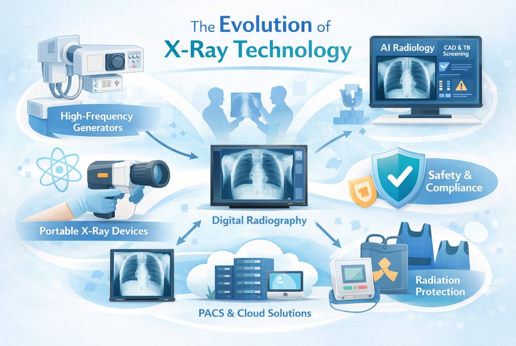
10 Innovations Transforming X-Ray Technology in 2026
The medical use of X-rays for diagnosis and treatment has proven to be immensely beneficial to society at large. Since the first dental X-ray was taken by Otto Walkhoff in 1896, the field has undergone a dramatic technological evolution aimed at reducing both occupational and patient exposure while maximizing diagnostic clarity. In the modern era, the industry is transitioning from conventional analog systems to high-frequency digital units and Artificial Intelligence (AI) powered diagnostics. These innovations are governed by rigorous standards, such as India's Atomic Energy Regulatory Board (AERB) Safety Codes and Central Drugs Standard Control Organization (CDSCO) medical device rules, to ensure safety remains paramount.
1. The Shift to High-Frequency Constant Potential Generators
A fundamental innovation in modern radiology is the replacement of conventional low-frequency systems with high-frequency (HF) constant potential generators.
Waveform Efficiency: Conventional X-ray units often produce a pulsating waveform that results in "soft" low-energy X-rays, which increase patient dose without aiding the diagnostic image.
Optimal Output: Modern HF generators (e.g., operating at 60-70 kVp) maintain a constant potential, ensuring the tube operates at peak voltage for the entire exposure.
Dose Optimization: This technology allows for shorter clinical exposure times—as low as 0.16 seconds for certain projections—effectively adhering to the ALARA (As Low As Reasonably Achievable) principle.
Filtration Standards: AERB mandates that equipment operating at constant potential must have a minimum total filtration of 2.5 mm Al equivalent to cut off useless radiation.
2. The Rise of Portable and Ultra-Portable (UP) Systems
Miniaturization has led to the development of X-ray units that provide high-resolution imaging in a compact form factor, suitable for field and humanitarian operations.
A. Defining Portability
The AERB defines Portable X-ray equipment as units intended to be carried by one or two persons, with a total weight not exceeding 12 kg. These are distinct from mobile units, which are moved on wheels.
B. Handheld Devices vs. Wall-Mounted Sources
Innovations such as the Nomad Pro handheld device have challenged the supremacy of traditional wall-mounted units.
Resolution Superiority: Independent research shows that handheld devices often provide significantly higher mean Line Pair (LP) resolution (6.05–6.55 lp/mm) compared to traditional wall-mounted sources (5.58–6.31 lp/mm).
Focal Spot Size: Handheld units often feature a smaller focal spot (0.4 mm) than fixed units (0.7 mm), which directly enhances the ability to resolve minute anatomical details.
User Preference: Clinical simulations indicate a 97% operator satisfaction rate for handheld units regarding ease of use, compared to 50% for conventional sources.
3. Digital Transformation: DR Panels and Advanced Detectors
The transition from analog film to Digital Radiography (DR) represents a landmark shift in how diagnostic information is captured and stored.
Direct Digital Capture: Modern systems utilize highly sensitive and dose-efficient LG Oxide Detectors (typically 14” x 17” panels) that produce an electrical signal immediately upon X-ray reception.
Computed Radiography (CR) Evolution: While CR uses Photostimulable Phosphor (PSP) plates that require scanning at resolutions such as 300 DPI, direct DR offers near-instantaneous visualization.
PACS and Cloud Integration: Modern radiology relies on Picture Archiving and Communication Systems (PACS) to store and distribute images without loss of data or quality. Recent implementations in India include cloud-based PACS to store scans for over three years.
4. AI-Powered Radiology and Computer-Aided Detection (CAD)
The most cutting-edge innovation in the market is the pairing of ultra-portable hardware with Artificial Intelligence (AI) and CAD software.
Automated Interpretation: CAD tools provide standardized interpretation of images, such as Chest X-rays (CXR), which is vital in regions where expert human readers are scarce.
Public Health Impact: This technology is currently the primary "hot" application for Tuberculosis (TB) screening in community-based settings. AI highlights the likelihood for a CXR to be suggestive of TB or other abnormalities, facilitating rapid triage.
Active Case Finding (ACF): Organizations like the William J. Clinton Foundation (WJCF) and India's Central TB Division utilize AI-integrated handheld units to bring high-spec diagnostics to the "doorstep" of symptomatic individuals.
5. Safety and Regulatory Design: The E-E-A-T Framework
Innovations are only as valuable as the safety frameworks that govern them. In India, X-ray Generating Equipment (XGE) is subject to strict oversight by the AERB and CDSCO.
A. Classification and Mandatory Registration
Effective January 1, 2021, all X-ray machines in India are classified as Class C (Moderate to High Risk) medical devices.
Regulatory Status: X-ray machines are legally considered "drugs" for regulatory purposes to prevent the proliferation of misbranded and inferior-quality products that lead to clinical misdiagnosis.
Approving Authority: CDSCO is the central authority for granting manufacturing and import licenses.
B. Built-in Design Safety
The AERB Safety Code SC-3 stipulates that radiation safety must be ensured "by design".
Leakage Radiation Limits: For general radiography, leakage through the tube housing must not exceed 1 mGy in one hour at 1.0 meter.
Safety Interlocks: Cabinet-type industrial and security equipment (e.g., XBIS or XRD) must feature fail-safe mechanisms and door interlocks that terminate X-ray emission if an access panel is opened.
Exposure Controls: Handheld devices must have proximity sensors to ensure they only fire when the object is at the correct distance and give clear audio/visual "ON" indications.
6. Operational Safety and the TDS Principle
Even with advanced technology, the human element of safety remains critical. All facilities must implement a Radiation Protection Programme (RPP).
Personnel Monitoring: All radiation workers must use Thermoluminescent Dosimeter (TLD) badges to track their cumulative dose. TLD badges must be worn below the lead apron at chest level.
Dose Limits: The annual effective dose limit for occupational workers is 20 mSv averaged over five years, while the limit for the public is 1 mSv.
The TDS Principle: Safety is maximized by optimizing Time, Distance, and Shielding.
Distance: For mobile/portable units, the operator should maintain at least 2.0 meters from the source.
Shielding: Use of lead aprons (0.25 mm lead eqv) and mobile protective barriers (1.5 mm lead eqv) is mandatory.
7. Quality Assurance (QA): The Compliance Baseline
To ensure that innovative equipment continues to perform accurately, periodic QA is a legal requirement.
Periodicity: End-users must ensure periodic QA is conducted by authorized agencies once every two years or after major repairs.
Critical QA Parameters:
kVp Accuracy: Within ± 5 kV.
Timer Accuracy: Percentage error not to exceed ± 10%.
Output Consistency: The Coefficient of Variation (CoV) must be < 0.05.
Alignment: Central beam alignment must be within < 1.5°.
Conclusion: The Future of Radiological Excellence
The latest innovations in X-ray technology—from high-frequency generators to AI-powered CAD software—are transforming radiology into a more precise, mobile, and patient-centric field. Success in this market is no longer defined by hardware alone, but by the integration of Expertise, Authoritativeness, and Trustworthiness (E-E-A-T) through rigorous compliance with AERB and CDSCO standards. As technology continues to evolve, the core objective remains constant: obtaining the optimum quality diagnostic information at the lowest possible radiation risk to the patient. By utilizing the e-LORA digital portal for licensing and maintaining religious adherence to Quality Assurance, India’s radiological community is well-positioned to lead the global shift toward safe and intelligent imaging.
About XRAYNEWS.NET
XRAYNEWS.NET provides updates on medical and industrial X-ray technologies, radiography insights, regulatory awareness, and imaging industry developments.
Regulatory Resources
© 2025. All rights reserved.
Video Resources
Popular Topics
Quick Links
Stay Updated with X-Ray Industry News
Subscribe to receive updates on X-ray technology developments, radiography insights, and industry news.