Digital Transformation in Radiology in India: DR Panels, PACS, Cloud Imaging & AI Trends
Explore how digital transformation is reshaping radiology in India through DR panels, PACS, cloud imaging, AI integration, and evolving AERB and CDSCO compliance standards.
INDUSTRY INSIGHTS & TECHNOLOGY
3/18/20264 min read
How DR Panels, PACS and Cloud Imaging Are Transforming Radiology
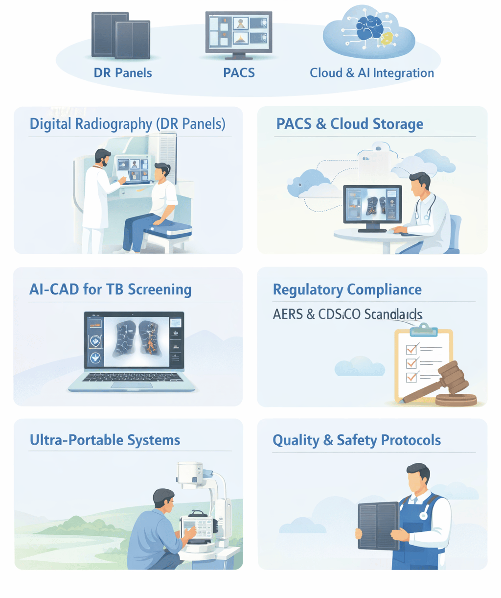
The Indian diagnostic imaging landscape is currently navigating a period of unprecedented digital evolution. Driven by strict regulatory mandates from the Atomic Energy Regulatory Board (AERB) and the Central Drugs Standard Control Organization (CDSCO), the industry has transitioned from traditional film-based systems to high-tech digital workflows. This transformation is anchored by three technological pillars: Digital Radiography (DR) Panels, Picture Archiving and Communication Systems (PACS), and Cloud-based Imaging.
The primary goal of this digital shift is the optimization of image quality and patient dose, adhering to the international ALARA (As Low As Reasonably Achievable) principle. By integrating Artificial Intelligence (AI) and cloud connectivity, India is bridging the diagnostic gap between urban centers and resource-limited community settings.
1. The Hardware Shift: DR Panels and the Direct Digital Advantage
The cornerstone of digital transformation is the Imaging Device, defined by the AERB as a detector unit or array of detectors that receives X-rays and produces an electrical or light signal.
A. Flat-Panel Detector Technology
Modern Digital Radiography (DR) systems utilize flat-panel detectors, such as high-performance LG Oxide Detectors (e.g., 14” x 17” panels). Unlike older technologies, DR provides near-instantaneous visualization of images on a portable workstation or laptop.
Dose Efficiency: DR detectors are "highly sensitive" and dose-efficient, allowing for superior image contrast with lower radiation levels.
Resolution Benchmarks: Digital systems provide high resolution; for instance, handheld digital devices often achieve significantly higher mean Line Pair (LP) resolution compared to traditional wall-mounted sources.
B. Transition from Computed Radiography (CR)
While many facilities still utilize Computed Radiography (CR), which uses Photostimulable Phosphor (PSP) imaging plates, the trend is moving toward direct DR.
Maintenance Needs: CR plates must be periodically evaluated for artifacts and cleaned. In contrast, digital detectors require periodic software calibration as per manufacturer recommendations to maintain performance.
2. Networking and Data Integrity: The Role of PACS
As imaging becomes entirely digital, the management of data becomes as critical as the acquisition of the image itself.
A. Defining PACS
A Picture Archiving and Communication System (PACS) is the digital infrastructure used to store, retrieve, and distribute radiological images. The AERB mandates that wherever PACS is used, facilities must ensure that:
The quality of patient images is maintained.
Patient information is not lost or unintentionally altered during storage or transmission.
B. Interconnectivity and Standards
Modern radiology departments rely on digital information systems interconnected by computer networks. According to AERB Safety Codes, these exchanges must be in accordance with national and international standards to ensure data fidelity. Procedures must be designed specifically to prevent any loss of diagnostic data.
3. The Rise of Cloud Imaging and AI Integration
The most recent trend in India is the migration of PACS to the Cloud, coupled with AI-powered Computer-Aided Detection (CAD).
A. Cloud-Based Storage
Cloud-based PACS allow for the storage of vast amounts of diagnostic data over several years (e.g., storing scans for a minimum of 3 years). This enables:
Telemedicine: Experts can read X-rays remotely, providing critical support in areas where expert human readers are scarce.
Accessibility: Community-based screening programs can upload images from the field directly to central health facilities.
B. AI-Powered CAD Software
Ultra-portable X-ray systems are increasingly paired with AI-CAD software, particularly for Tuberculosis (TB) screening.
Automated Interpretation: CAD tools provide standardized interpretation by highlighting the likelihood of abnormalities (like TB) in a Chest X-ray (CXR).
Triage Efficiency: AI highlights presumptive cases for molecular testing, making high-throughput screening feasible in resource-limited conditions.
4. Regulatory Framework: CDSCO and Class C Classification
A pivotal moment for the Indian X-ray market occurred on January 1, 2021, when X-ray machines were officially classified as Class C (Moderate to High Risk) medical devices.
Mandatory Registration: Under the Medical Device Rules 2017, all manufacturers and importers must register their X-ray equipment with the CDSCO.
Prevention of Misdiagnosis: This regulation was implemented to curb the spurt of misbranded or inferior-quality products that lead to inaccurate diagnostic reporting.
Registration Fees: Importers face a structured fee system, including $3,000 for manufacturing plant registration and $1,500 per product.
5. Operational Compliance via the e-LORA Portal
The e-LORA (e-Licensing of Radiation Applications) system is the mandatory digital gateway for all AERB regulatory transactions in India.
A. The Procurement Pathway
Before acquiring a digital X-ray unit, the Employer of a facility must obtain a Procurement Letter via e-LORA.
Requirement: The facility must select an AERB Type Approved model and an authorized supplier.
Approval: Procurement approval usually takes only a few hours once the digital application is submitted.
B. Licensing for Operation
No digital equipment can be legally used for patient diagnosis until an Operational Licence is granted.
Pre-requisites: This requires uploading an Installation Report and a Quality Assurance (QA) Report.
Approval Duration: The AERB typically issues the license within 30 days of a successful application.
6. Safety Protocols and Quality Assurance (QA)
Digital transformation does not replace the need for traditional radiation safety; rather, it enhances it through better monitoring and performance checks.
A. Mandatory QA Tests
The end-user is responsible for ensuring that periodic QA is carried out by authorized agencies every two years. Critical digital parameters tested include:
Operating Potential (kVp) Accuracy: Within ± 5 kV.
Output Consistency (CoV): Must be < 0.05 to ensure stable imaging.
Beam Alignment: Central beam alignment must be < 1.5° to prevent image distortion.
B. Personnel Safety Tools
Regardless of how advanced the digital system is, the Time-Distance-Shielding (TDS) principle remains essential.
TLD Badges: All radiation workers must use Thermoluminescent Dosimeter (TLD) badges, typically worn below the lead apron at chest level.
Lead Equivalence: Protective aprons must have a minimum of 0.25 mm lead equivalence, while mobile barriers require 1.5 mm.
7. Trends in Field Applications: Ultra-Portable Systems
The rise of Ultra-portable (UP) systems (weighing under 12 kg) represents the "hot" product trend in India’s digital transformation.
Field Utility: These battery-powered units are ideal for community Active Case Finding (ACF) camps where conventional infrastructure is unavailable.
Safety in the Field: For portable security scanners, the total dose at 30 meters should not exceed 5 µSv per scan.
Operator Distance: In field operations, radiographers should maintain a minimum distance of 2 meters from the source during exposure.
Conclusion
Digital transformation in Indian radiology is far more than a hardware upgrade; it is a holistic shift toward a safe, integrated, and high-quality diagnostic ecosystem. By transitioning to DR Panels, leveraging Cloud-based PACS, and adopting AI-CAD interpretation, Indian healthcare providers are meeting global standards of diagnostic excellence.
However, the foundation of this progress remains a deep commitment to AERB and CDSCO regulatory compliance. Success in this digital era is defined by religious adherence to biennial Quality Assurance, meticulous e-LORA licensing, and a culture of safety that ensures every digital image is obtained at the lowest possible radiation risk to the patient. For clinics and hospitals, staying ahead of these digital trends is not just a technological advantage—it is a legal and ethical mandate to provide the best possible care to the public.
About XRAYNEWS.NET
XRAYNEWS.NET provides updates on medical and industrial X-ray technologies, radiography insights, regulatory awareness, and imaging industry developments.
Regulatory Resources
© 2025. All rights reserved.
Video Resources
Popular Topics
Quick Links
Stay Updated with X-Ray Industry News
Subscribe to receive updates on X-ray technology developments, radiography insights, and industry news.