
What is an X-ray? How It Works & Why It's Used in Medicine
Learn how X-rays work, their medical uses, safety, and the difference between digital and traditional X-rays. Easy guide for patients and students.
5/1/20255 min read
Our modern age of medicine, X-rays are most prevalent and superior machine utilized to scan inside the human body without needing to open them up. To diagnose a fracture in a bone, diagnose pneumonia inside the lungs, diagnose tumors when too advanced to repair, X-rays provide doctors a speedy, safe, and certain method of diagnosing.
But what is an X-ray, and how does it work, and why is it so integral to medicine today?
In this special report for X-ray News, we'll break it down in plain language so you can see not only the science behind it, but just how pervasive X-rays are a part of daily medicine.
What is an X-ray?
An X-ray is an electromagnetic radiation. X-rays propagate in waves, just like visible light, radio waves, or microwaves. X-rays do indeed have so much more energy than visible light, and precisely for that, can penetrate soft body tissues.
German physicist Wilhelm Conrad Roentgen discovered X-rays in 1895, which revolutionized the field of medical science. Roentgen discovered that X-rays pass easily through materials such as cardboard and tissue but not materials such as bone and metal. It gave him the first Nobel Prize in Physics in 1901.
Now let us move step by step how the process of working of X-ray imaging is done:
1. X-ray Production
There is an X-ray machine which has an X-ray tube unit. Within the tube, electrons are given extremely high speeds in the direction of a metal target (tungsten is commonly used). When they impact electrons within the metal, X-rays are generated.
2. The Radiation Travels through the Body
The X-rays are pointed at some part of the patient. As they travel through, they are stopped by some tissues to some extent
- Bones (compact tissue) stop a lot of the X-rays and colored as white.
- Muscles and organs stop less and colored as gray.
- Air cavities such as lungs pass through to pass on most of their X-rays and colored as black.
3. Recording the image
The X-rays are stored on a film or digital detector for free. This creates a shadow image of the inside structure, which can be analyzed by doctors.
X-ray Imaging Techniques Types:
1. Conventional X-ray (Radiography)
X-ray imaging of chest, bone, and abdominal. Outputs black-and-white images in 2D.
2. Fluoroscopy
Creates moving X-ray images, which are usually utilized for procedures and surgery like angiograms.
3. Computed Tomography (CT Scan)
A CT scan scans multiple X-ray pictures at various angles and constructs 3D imaging. It allows superimposing an image of cross-sectional tissue and organ information.
4. Dental X-rays
For the diagnosis of cavities, bone loss, and tooth misalignment.
X-rays are used in intra-nearly-all areas of medical specializations. Some of the very frequent uses are:
Diagnosis of Fracture and Dislocation
- X-rays are routine to detect joint disease or fractured bone due to arthritis.
Chest X-ray
- For diagnosing tuberculosis or pneumonia, lung cancer, or lung congestion.
Dental X-rays
- They reveal cavities, issues with wisdom teeth, and jawbone development.
Mammography
- Initial X-ray for breast cancer screening.
- Spinal and Skeletal Exams
- To detect scoliosis, arthritis, and other skeletal conditions.
- Abdominal X-rays
- To expose kidney stones, blockage, or something ingested.
⚠️ Is X-ray Safe?
No one gets scared with the word radiation, but diagnostic X-rays contain very little radiation. The amount is so tiny that it won't hurt most people, much less if X-rays are repeated and repeated and repeated again and again and again for a long, long time.Repetition or exposure, however, must be reduced to bare minimum. Medical professionals thus follow the ALARA principle – "As Low As Reasonably Achievable."
Precautions while X-raying
- Always inform your doctor if pregnant.
- Use thyroid shields or lead aprons during imaging.
- Use digital X-rays, 70–80% less radiation than film X-rays.
Digital X-rays or Film X-rays
Science of X-ray has evolved a long way from the good old days. X-rays, or film X-ray, once had a picture on photographic film. In fact, the process is chemical, it is time consuming, and after knowing the process, expose the patient to more radiation. Digital X-rays did stop this trend, however. Digital X-rays never use film but digital sensors instead, to produce images instantly on a screen monitor. Digital X-rays provide improved images, reduced radiation dose, and silent electronic storage of electronic medical records electronically. Digital X-rays are easy to correct and enlarge for better diagnosis and can be e-mailed instantaneously or archived by cloud storage, best suited for telemedicine and remote consultation. Due to such benefits, digital radiography is the norm in all diagnostic clinics and hospitals today.
All thanks to R&D, X-rays are no longer the boring dry grey bone scans anymore. Some of the nice developments are:
1. Portable X-ray Machines
Battery-powered light machines enable imaging at home, in an ambulance, or in a remote clinic.
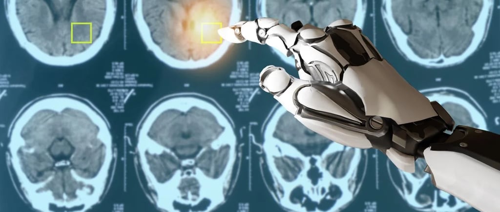
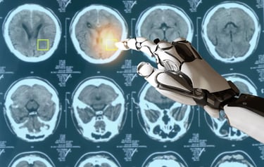
2. Artificial Intelligence in Radiology
Artificial Intelligence can identify lung cancer, fractures, and TB on an X-ray faster and better than the naked eye sometimes.
3. Cone Beam CT (CBCT
Used in ENT clinics and dental clinics to produce 3D images with reduced radiation exposure.
4. Wireless Digital Detectors
Allows real-time sharing and cloud storage of X-rays for tele-radiology (telemedicine diagnosis).
Who Does Xrays?
A trained radiologic technologist (radiographer) will usually take the X-ray, and a radiologist (physician trained in imaging) will interpret the image and report back on it to the referring physician.
Advantages of X-ray imaging
- No invasion and no pain
- Universal and quick
- Cheap
- Accurate for chest and bone diagnosis to a great extent
- Critical when it comes to detection of disease at an early stage
When You Would Get an X-ray? Your doctor would recommend an X-ray if:
- You suffer from trauma or fracture, traumatic infection, broken joint or bone
- You present signs and symptoms of infection or respiratory disease
- You have incontinence urinary, swelling, or GI symptoms
- Need chronic disease monitoring like arthritis
Always adhere to your doctor's instructions and never postpone with imaging, if ordered.
Explanation: Essential for early diagnosis of the disease
More than a century ago, X-rays were already saving lives. From a broken bone to a life-threatening disease such as cancer, they assist physicians in taking an accurate picture of what is occurring within the body — and doing it fast and without harm.
Thanks to advances in digital technology, portable equipment, and computer-aided diagnoses, X-ray imaging is more accurate, quicker, and more routine.
Physician, physician assistant, medical student, or medical tech enthusiast, X-ray equipment tour into the eyes of one of medicine's greatest modern gadgets.
Here are suggested references you can include at the end of your blog. These are general references appropriate for a medical explainer article and based on reliable sources:
References
Radiological Society of North America (RSNA). What is an X-ray (Radiograph)?
https://www.radiologyinfo.org/en/info/chestradWorld Health Organization (WHO). Radiation: X-rays
https://www.who.int/news-room/questions-and-answers/item/radiation-x-raysNational Institute of Biomedical Imaging and Bioengineering (NIBIB). X-ray Imaging
https://www.nibib.nih.gov/science-education/science-topics/x-ray-imagingU.S. Food & Drug Administration (FDA). Medical X-ray Imaging
https://www.fda.gov/radiation-emitting-products/medical-x-ray-imagingMayo Clinic. X-ray
https://www.mayoclinic.org/tests-procedures/x-ray/about/pac-20395303American College of Radiology (ACR). ACR Appropriateness Criteria®
https://www.acr.org/Clinical-Resources/ACR-Appropriateness-CriteriaRoentgen, W. C. (1895). On a New Kind of Rays (original publication of X-ray discovery).
https://www.ndsu.edu/pubweb/~mcclean/plsc431/radiation/xray.htm