
Understanding the Power of X-Rays: A Beginner’s Guide
X-rays have been one of the most revolutionary medical technologies in history. From their discovery over a century ago, they have transformed the way we diagnose and treat various health conditions. In this beginner’s guide, we’ll explore the science behind X-rays, their applications, and how they continue to shape the medical field today.
3/5/20253 min read
What are X-rays?
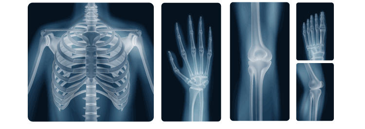
X-rays are a form of electromagnetic radiation, just like visible light, but with much higher energy. These waves are capable of passing through the human body, and this unique ability makes them incredibly valuable in medical imaging.
When X-rays pass through the body, they are absorbed at different rates by various tissues. Dense tissues like bones absorb more X-rays, while less dense tissues like muscles or organs absorb fewer. This variation creates a contrast in the resulting image, allowing medical professionals to see the inside of the body without invasive procedures.
The Discovery of X-Rays
The discovery of X-rays is credited to German physicist Wilhelm Conrad Roentgen in 1895. Roentgen was conducting experiments with cathode rays when he noticed that a fluorescent screen in his laboratory began to glow without being in direct contact with the cathode rays. He realized that a new type of radiation, which he called "X-rays," was responsible for the phenomenon.
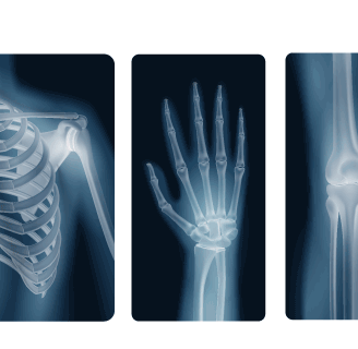
Shortly after the discovery, Roentgen used X-rays to capture the first image of a human hand, complete with its bones. This marked the beginning of a new era in medical diagnostics.
How Do X-Rays Work?
X-ray machines work by directing a controlled beam of X-rays onto the body. The X-rays then pass through the body and strike a detector on the opposite side. Depending on the density of the tissues, some X-rays are absorbed, while others pass through and hit the detector. The information gathered is then processed into an image, which is displayed on a screen for analysis.
Key components of an X-ray system:
X-ray Tube: This is where the X-rays are produced. The tube contains a cathode and an anode that generate the X-rays when high-energy electricity passes through them.
Detector: This captures the X-rays that pass through the body and converts them into a digital image.
Control Panel: This allows the operator to adjust the settings, such as the intensity of the X-ray beam, to ensure optimal image quality.
The Different Types of X-Ray Imaging
X-rays are used in various types of imaging techniques, each serving a specific purpose in the diagnosis and treatment of medical conditions. Here are the most common types:
Traditional X-Ray (Radiography)
This is the most basic form of X-ray imaging, where a single X-ray beam is used to capture an image of the body. It’s commonly used to detect fractures, infections, and certain diseases, like pneumonia.Computed Tomography (CT) Scan
A CT scan is an advanced form of X-ray imaging that uses multiple X-ray images taken from different angles to create detailed cross-sectional images of the body. CT scans are particularly useful for detecting cancers, internal injuries, and complex bone fractures.Mammography
Mammograms are specialized X-ray images of the breast, used primarily for the early detection of breast cancer. The process involves compressing the breast between two plates to get clear images.Fluoroscopy
Fluoroscopy allows doctors to view moving images of the inside of the body in real time. It’s often used during surgeries, in the placement of catheters, or to examine the digestive system.Dental X-Rays
These are small X-ray images that focus specifically on the teeth and jaw. Dental X-rays are used to detect cavities, infections, bone loss, and other dental conditions.
The Benefits of X-Ray Technology
Non-Invasive
One of the primary benefits of X-ray imaging is that it is non-invasive, meaning it does not require surgery or other invasive procedures to get a detailed look inside the body. This makes it a safer and less traumatic alternative to diagnostic methods that require incisions.Speed and Convenience
X-ray images can be produced quickly, making them ideal for emergency situations where time is of the essence. In addition, digital X-ray systems allow for instant image viewing, speeding up the diagnostic process.Cost-Effective
Compared to other imaging technologies like MRI or ultrasound, X-rays are relatively inexpensive, making them a widely accessible diagnostic tool in hospitals and clinics worldwide.Diagnostic Clarity
X-ray images offer detailed insights into bones and certain tissues, making it easier to identify fractures, infections, and abnormalities in the body. This clarity aids in accurate diagnosis and treatment planning.
Safety of X-Rays
While X-rays are generally considered safe, they do involve exposure to a small amount of ionizing radiation. This is why they are usually only performed when necessary, and the amount of radiation is carefully controlled to minimize any potential risks.
For patients who require frequent X-ray imaging, modern technology, such as digital X-rays, has drastically reduced radiation exposure compared to older methods. Additionally, special precautions are taken for pregnant women and young children, who are more sensitive to radiation.


Reference Website Link:
American College of Radiology (ACR)
Website: https://www.acr.org/
Website: https://www.radiologyinfo.org/
National Institutes of Health (NIH) – National Library of Medicine (MedlinePlus)
Website: https://medlineplus.gov/
Mayo Clinic
Website: https://www.mayoclinic.org/
World Health Organization (WHO) – Radiation Safety
Website: https://www.who.int/