
The Evolution of X-ray Imaging: From Film to Digital
X-ray imaging is one of the most significant breakthroughs in the history of medical technology. It revolutionized how doctors diagnose and treat patients, enabling them to peer inside the human body without making a single incision. However, what many people may not realize is how much X-ray technology has evolved over the years. From its early beginnings with film-based X-ray systems to the cutting-edge digital imaging techniques used today, this technology has come a long way. Let’s take a deep dive into the evolution of X-ray imaging and explore how it has transformed medical diagnostics.
3/7/20254 min read
1. The Birth of X-ray Technology
X-ray technology was discovered by Wilhelm Conrad Roentgen in 1895, a groundbreaking event that earned him the first Nobel Prize in Physics in 1901. Roentgen’s discovery opened the doors to non-invasive diagnostic imaging, as he was the first to show that X-rays could pass through the human body and produce an image of its internal structures on a photographic plate.
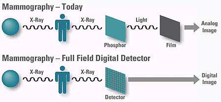
For several decades after the discovery, X-ray imaging primarily used film-based methods to capture the images. This analog process involved shining X-rays through the body and onto a photographic plate, which would then develop the image after exposure. The film was sensitive to radiation, which created the visual representation of bones and organs inside the body.
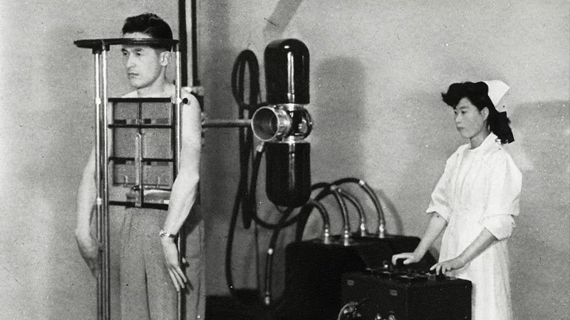
2. Film-Based X-ray Systems
In the mid-20th century, film-based X-ray technology became widely used in medical settings. Hospitals and diagnostic centers would use large machines to expose patients to X-rays, and the resulting film would be developed in a darkroom. These films were the first true X-ray images, allowing doctors to examine fractures, detect infections, and even diagnose serious conditions like tuberculosis.
However, while film-based X-rays were a revolutionary development, they came with a set of limitations. The process was slow and required a significant amount of manual labor, from developing the film to storing large physical archives of images. Additionally, the quality of the images was not always perfect, with issues like graininess or exposure problems making some X-rays difficult to read.
3. The Introduction of Digital X-ray Imaging
By the late 20th century, the limitations of traditional film-based X-ray systems prompted a shift towards digital technology. In the 1980s, the medical community began experimenting with digital X-rays, leading to the development of the first digital radiography systems in the early 1990s.
Digital X-ray imaging works by converting X-ray data into digital signals that can be displayed on a computer screen. This technology eliminates the need for film and chemicals used in the traditional film-based process. Instead, the X-rays are captured by a digital detector, which then converts them into an electronic image. This image can be immediately viewed, edited, and stored digitally, significantly speeding up the diagnostic process and making it easier to share and store images.
4. Benefits of Digital X-rays
The transition from film-based X-rays to digital imaging brought a host of benefits that have transformed medical imaging practices:
- Immediate Results
One of the key advantages of digital X-ray imaging is that it provides immediate results. Instead of waiting for films to develop, doctors and radiologists can view digital images almost instantly. This speeds up the diagnostic process and allows for quicker decision-making, which can be critical in emergencies.
- Enhanced Image Quality
Digital X-rays offer better image quality compared to film-based systems. With digital sensors, radiologists can adjust the contrast, brightness, and resolution of images, allowing them to see finer details that might be missed in traditional X-rays. This increased precision improves diagnostic accuracy.
- Lower Radiation Exposure
Digital X-ray systems use less radiation than film-based systems to produce clear images. This reduction in radiation exposure is important for patient safety, particularly for those requiring multiple X-ray exams, such as cancer patients or individuals with chronic conditions.
- Storage and Sharing
Digital images can be stored in electronic databases and shared quickly with other medical professionals. This has made collaborating on patient cases easier, as doctors in different locations can access and review images without waiting for physical films to be sent by mail or courier.
- Environmental Impact
With digital imaging, the need for chemical developers and physical film prints is eliminated, reducing the environmental impact of X-ray procedures. This eco-friendly aspect has made digital X-rays a more sustainable option for medical centers.
5. The Rise of 3D and Advanced Imaging
In recent years, digital X-ray imaging has continued to evolve with the advent of more advanced technologies. One of the most notable developments has been the introduction of 3D X-ray imaging and tomosynthesis. These innovations allow for the creation of three-dimensional images from a series of two-dimensional X-ray slices.
3D X-ray technology is particularly useful in dental imaging, where it helps to visualize the teeth, jaw, and surrounding structures in more detail than traditional 2D X-rays. This technology is also increasingly being used in other specialties, such as breast imaging, where digital breast tomosynthesis has improved early detection of breast cancer by providing clearer images of dense breast tissue.
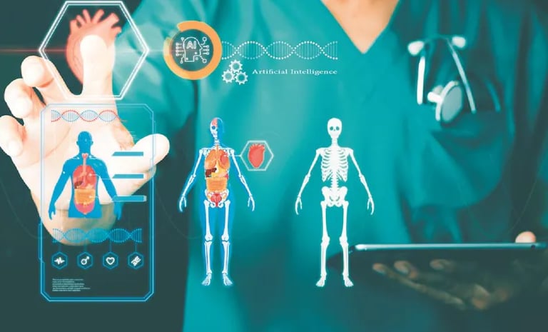
6. Artificial Intelligence in X-ray Imaging
The most recent frontier in X-ray technology involves the integration of artificial intelligence (AI) and machine learning. AI algorithms are being used to help analyze and interpret X-ray images faster and with greater accuracy. These systems can detect patterns that might be too subtle for the human eye, such as early signs of diseases like cancer, pneumonia, or fractures.
AI-powered X-ray systems can also assist radiologists by automatically flagging potential areas of concern, making the diagnostic process even more efficient. As AI continues to advance, it is expected to further transform the way X-ray imaging is used in medical practice, improving both speed and accuracy.
7. Looking Ahead: The Future of X-ray Imaging
The journey from film to digital imaging has dramatically enhanced the way doctors and radiologists approach diagnosis and treatment. Today, digital X-ray technology is faster, safer, and more efficient than ever before, and the integration of 3D imaging and AI has only expanded its capabilities.
Looking ahead, there are even more exciting developments on the horizon. Researchers are exploring the potential of quantum imaging and nanotechnology to create X-ray systems that are even more powerful, with the ability to capture images at the molecular level. Such advancements could enable earlier diagnosis of diseases and further refine treatments, ultimately improving patient care and outcomes.
Reference Website Link:
Radiological Society of North America (RSNA)
RSNA - Radiology News
National Institute of Biomedical Imaging and Bioengineering (NIBIB)
NIBIB - Medical Imaging
American College of Radiology (ACR)
ACR - X-ray Technology
PubMed Central (PMC) - National Library of Medicine
PubMed Central - X-ray Imaging
World Health Organization (WHO)
WHO - Medical Imaging