
How X-Rays Are Revolutionizing Medical Diagnostics
1. Enhanced Diagnostic Accuracy with Advanced Imaging
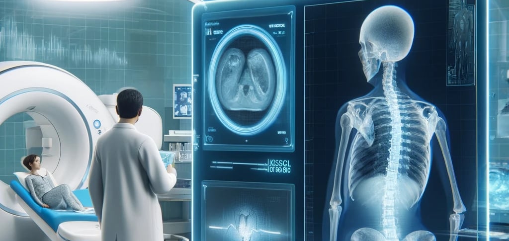
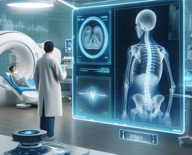
One of the most significant advancements in X-ray technology is the ability to produce high-resolution images that provide an unparalleled level of detail. In modern diagnostic radiology, traditional X-rays have been enhanced with digital imaging and advanced detectors, leading to sharper images that allow doctors to detect even the smallest abnormalities.
For example, digital X-ray imaging enables radiologists to see fine bone fractures, early signs of osteoporosis, and even subtle tumors that might have been missed with older technology. Digital systems also improve contrast, making it easier to distinguish between different tissues, such as healthy organs and diseased areas.
2. Speed and Efficiency in Diagnosis
Traditional X-ray machines required film development, which could take several minutes, or even hours, to process. Today, with digital radiography, images are available almost immediately after the scan is taken. This dramatically reduces wait times and allows doctors to quickly assess the images, leading to faster diagnoses and treatment plans.
Faster imaging also improves emergency care. For example, in trauma situations, doctors can obtain quick X-ray results, such as detecting broken bones or internal injuries, which can be life-saving in critical moments.
3. The Rise of 3D Imaging and CT Scans
CT scans (computed tomography) have taken traditional X-rays to the next level by combining X-ray technology with computer processing to create 3D images of the body. This technology allows doctors to view cross-sections of organs, bones, and tissues, providing more in-depth insights into a patient’s condition.
For example, CT scans are invaluable in diagnosing complex issues such as brain injuries, abdominal diseases, and certain types of cancers. By offering a clearer, multi-dimensional view of the body, CT scans allow healthcare professionals to make more informed decisions regarding treatment options.
4. X-Rays in Early Detection of Diseases
Early detection is crucial in treating many diseases, and X-ray technology plays a key role in identifying conditions at their earliest stages. One of the most well-known examples is mammography, a specialized X-ray technique used to detect breast cancer. Regular mammograms can help catch tumors long before they become palpable or symptomatic, increasing survival rates through early intervention.
X-rays also play a vital role in detecting lung diseases, such as pneumonia or tuberculosis, as well as identifying heart conditions like congestive heart failure. As the technology continues to improve, doctors are able to spot even the faintest signs of disease, leading to faster treatment and better outcomes for patients.
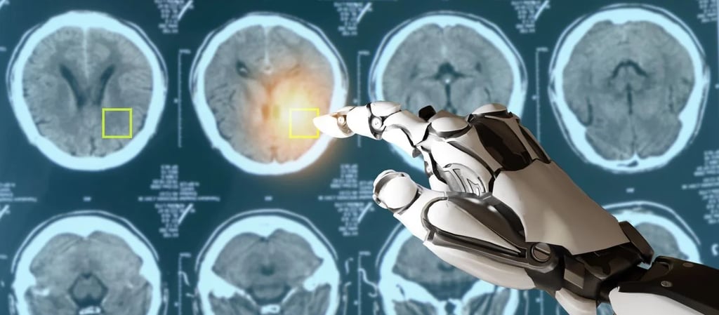
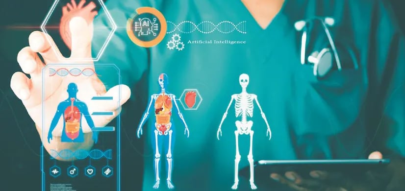
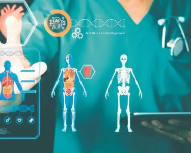
5. The Integration of Artificial Intelligence (AI) with X-Ray Technology
In recent years, AI has made its way into the world of radiology, significantly improving the capabilities of X-ray imaging. Machine learning algorithms are being integrated into radiology systems to help identify patterns and anomalies in X-ray images.
AI-powered tools can analyze large volumes of X-ray data quickly, highlighting areas that may require further investigation. This can help reduce human error and increase the efficiency of diagnosing conditions, especially in busy medical settings. For instance, AI is being used to help radiologists detect early-stage lung cancer, fractures, and even spinal abnormalities that might be missed by the human eye.
Moreover, AI’s ability to learn from large datasets means that its diagnostic capabilities continue to improve over time, potentially leading to even higher accuracy rates in the future.
6. Minimizing Radiation Exposure for Patients
One concern with traditional X-rays has always been the risk of radiation exposure. While X-rays are generally safe when used properly, the increasing focus on patient safety has led to innovations that minimize radiation exposure. For instance, new low-dose X-ray technology significantly reduces the amount of radiation needed for imaging while still delivering high-quality results.
Moreover, fluoroscopy, a real-time X-ray imaging technique, has evolved to offer enhanced control over radiation dose delivery. With these advances, doctors can ensure that patients receive the necessary diagnostic information without excessive exposure to radiation.


7. Expanding Access to X-Ray Technology
X-rays are no longer limited to major hospitals and clinics. Thanks to portable X-ray machines, healthcare providers can bring diagnostic imaging to patients in remote or underserved areas. This is especially important in emergency settings, rural communities, or for patients who are bedridden and unable to visit a clinic.
Portable X-ray machines are lightweight and easy to operate, enabling healthcare workers to deliver accurate diagnoses on-site. This capability is particularly useful in emergency rooms, ambulances, and field hospitals, where quick diagnostic information can be crucial for patient care.
8. Personalized Medicine and Treatment
One of the most exciting developments in modern medicine is the shift towards personalized treatments. X-ray technology is helping doctors tailor treatments based on a patient’s unique anatomy and condition. By using advanced imaging techniques like 3D mammography or dual-energy X-ray absorptiometry (DEXA), physicians can design more effective treatment plans that are specifically suited to the patient’s needs.
For example, in orthopedic medicine, X-rays are used to assess bone density and the severity of conditions like osteoporosis, allowing for personalized medication regimens and physical therapy plans that maximize the patient’s health outcomes.
Reference Website Link:
Radiological Society of North America (RSNA)
Link: https://www.rsna.org
National Institutes of Health (NIH)
Link: https://www.nih.gov
American College of Radiology (ACR)
Link: https://www.acr.org
Health Physics Society
Link: https://hps.org
Science Direct – X-ray Imaging in Medicine