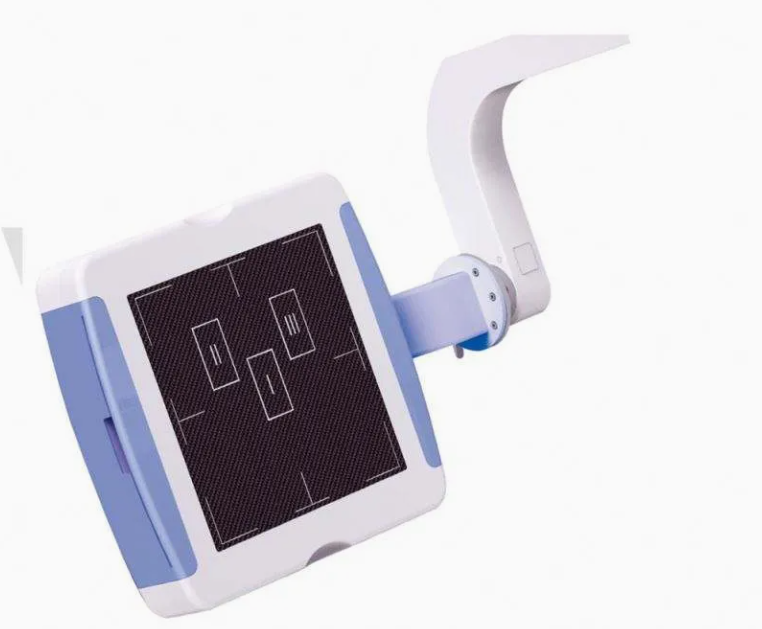
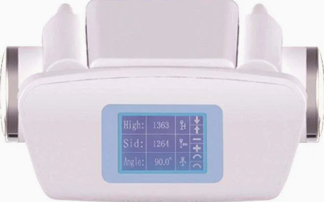
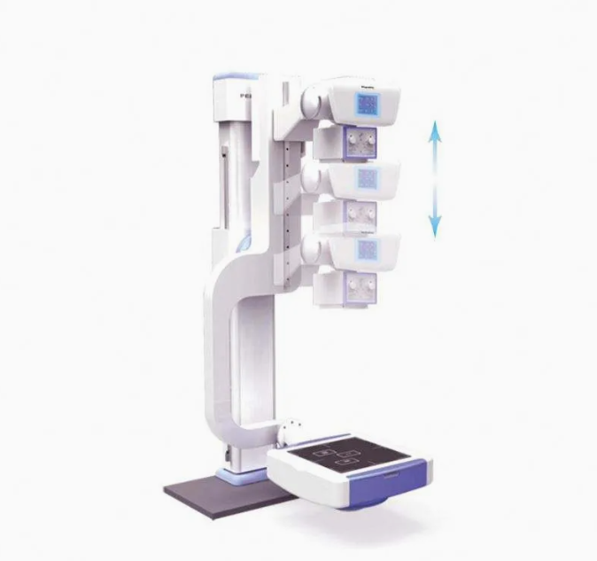
Radiology Digital 500mA Medical X Ray Machine
The Radiology Digital 500mA Medical X-ray Machine is an advanced diagnostic tool used in medical imaging to produce high-quality X-ray images for a variety of applications. With a 500mA output, this machine delivers high-resolution images, making it suitable for a wide range of radiological examinations. It uses digital radiography (DR) technology, which provides superior image quality, faster processing times, and lower radiation exposure compared to traditional film-based systems.
This type of X-ray system is commonly used in hospitals, diagnostic centers, and clinics, where detailed and clear imaging is crucial for accurate diagnosis and treatment planning. The combination of high mA (500mA) power output and digital imaging technology ensures that even dense tissues like bones, organs, and soft tissues can be imaged clearly with minimal exposure.
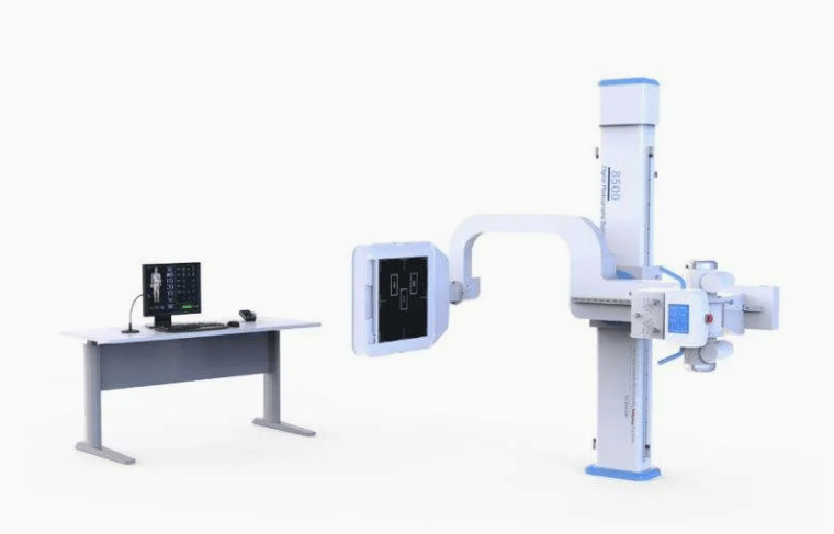

Key Features:
500mA High Power Output:
The 500mA current provides powerful X-ray generation, which is ideal for capturing high-resolution images, particularly for thick or dense body structures like bones and organs. This high output ensures accurate imaging for a wide range of patients, from those with larger body types to complex diagnostic cases.
Digital Radiography (DR) Technology:
The machine uses digital radiography (DR), which enables the production of instant, high-quality images without the need for film or chemical processing. This technology also allows for easier storage, retrieval, and sharing of images via digital networks.
High-Resolution Imaging:
The 500mA output paired with DR technology allows the machine to produce sharp, detailed images. This ensures that even the most challenging diagnostic cases, such as bone fractures or soft tissue abnormalities, can be examined accurately.
Automated Exposure Control:
Many modern systems come with automatic exposure control (AEC), which automatically adjusts exposure settings based on the patient's size and the area being imaged. This feature helps ensure consistent image quality while minimizing unnecessary radiation exposure.
User-Friendly Interface:
Digital systems often include intuitive controls or touchscreen interfaces that simplify the operation of the machine. Healthcare providers can easily adjust settings such as exposure time, voltage, and current to suit specific diagnostic needs.
Reduced Radiation Exposure:
Digital radiography systems are designed to reduce the amount of radiation needed to obtain high-quality images, which is safer for both patients and healthcare providers.
Quick Image Processing:
DR technology allows for rapid image processing, providing near-instant results. This speeds up diagnosis, enables faster treatment planning, and improves patient throughput in busy clinical environments.
Built-in Safety Features:
Advanced safety features like dose monitoring, automatic shielding, and radiation protection ensure the system complies with regulatory standards, ensuring the safety of both the patient and healthcare personnel.
Remote Access and Integration:
Many digital X-ray systems allow for integration with hospital information systems (HIS) or picture archiving and communication systems (PACS), enabling remote access to images for consultation and sharing among healthcare professionals.
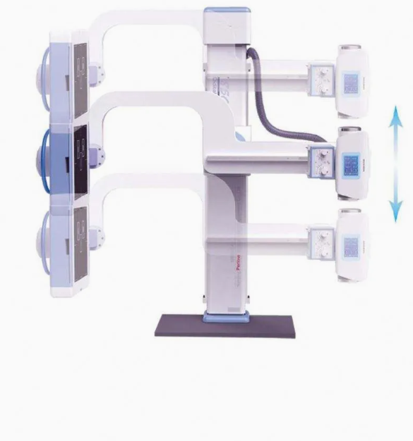

Applications:
General Radiography:
The 500mA X-ray machine is commonly used for standard diagnostic imaging, such as chest X-rays, abdominal X-rays, and skeletal imaging. It is ideal for routine screening and diagnostic purposes.
Orthopedics:
It is used for diagnosing bone fractures, joint problems, arthritis, and other musculoskeletal conditions. The high power output ensures that bone structures are clearly visualized, even in dense or complex cases.
Emergency Medicine:
In emergency settings, rapid and accurate imaging is required. This X-ray system is highly effective in diagnosing trauma-related injuries like fractures, dislocations, and internal bleeding, providing quick results in critical care situations.
Cardiology:
It can be used for imaging the heart and lungs to diagnose conditions such as heart disease, lung infections, and pulmonary edema. The clarity of images provided by the system helps in the accurate evaluation of the chest area.
Pediatrics:
Pediatric patients benefit from the lower radiation exposure offered by the digital system, while still ensuring high-quality images for the diagnosis of conditions like pneumonia, bone fractures, or congenital abnormalities.
Dental Imaging:
Although not primarily designed for dentistry, the machine may also be used for full-body or panoramic dental X-rays, especially in more advanced diagnostic centers.
Pulmonary Imaging:
The system is excellent for capturing detailed chest images, helping diagnose lung diseases, infections, and conditions such as tuberculosis, pneumonia, or lung cancer.

Why Choose a Radiology Digital 500mA Medical X-ray Machine?
High-Quality Imaging:
The 500mA power output ensures that the machine can produce detailed, high-resolution images, which are essential for accurate diagnosis, particularly in dense tissue areas such as bones and organs.
Faster Diagnosis:
With digital radiography, images are processed almost instantaneously, speeding up the diagnostic process and enabling quicker decision-making for treatment.
Lower Radiation Exposure:
Digital X-ray machines use advanced technologies that require lower radiation doses compared to conventional film-based systems, improving patient safety and reducing the risks associated with radiation exposure.
Cost-Effectiveness:
The initial investment in a digital X-ray machine may be higher than film-based systems, but the savings on film, chemicals, and labor costs over time make it a more cost-effective solution in the long term.
Ease of Storage and Sharing:
Digital images can be easily stored in electronic databases, reducing physical storage space requirements and enabling easier sharing and retrieval of images for consultations or second opinions.
Improved Workflow Efficiency:
The rapid image acquisition and processing speed improve the overall workflow, allowing for more patients to be seen and examined in less time, which is critical in busy healthcare environments.
Advanced Safety Features:
With built-in dose reduction technology, automatic exposure control, and other safety features, the system ensures that radiation exposure is kept to a minimum without compromising on image quality.
Remote Accessibility:
The system's integration with PACS and HIS allows healthcare providers to remotely access images, enabling telemedicine and faster consultation with specialists, improving the overall care process.
Reliability and Durability:
Digital 500mA X-ray machines are designed to operate reliably in high-volume environments, making them a solid investment for hospitals and clinics that require consistent, long-term performance.
