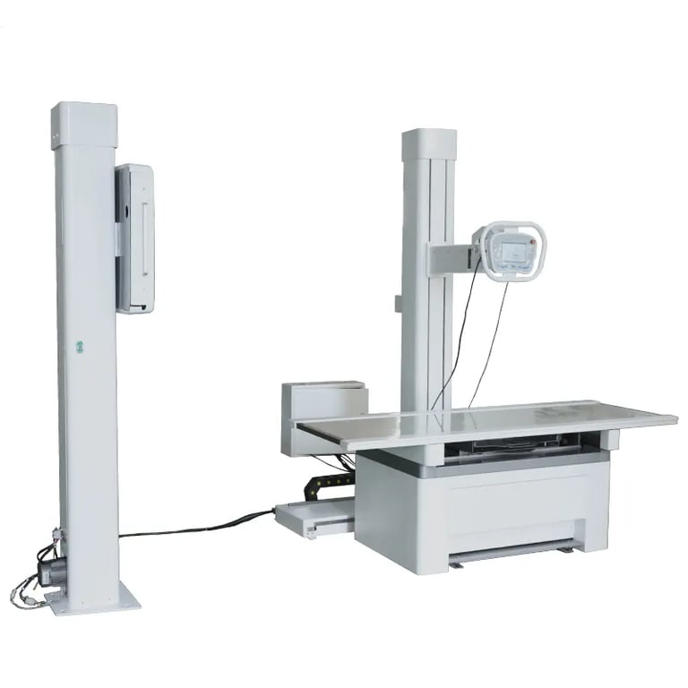
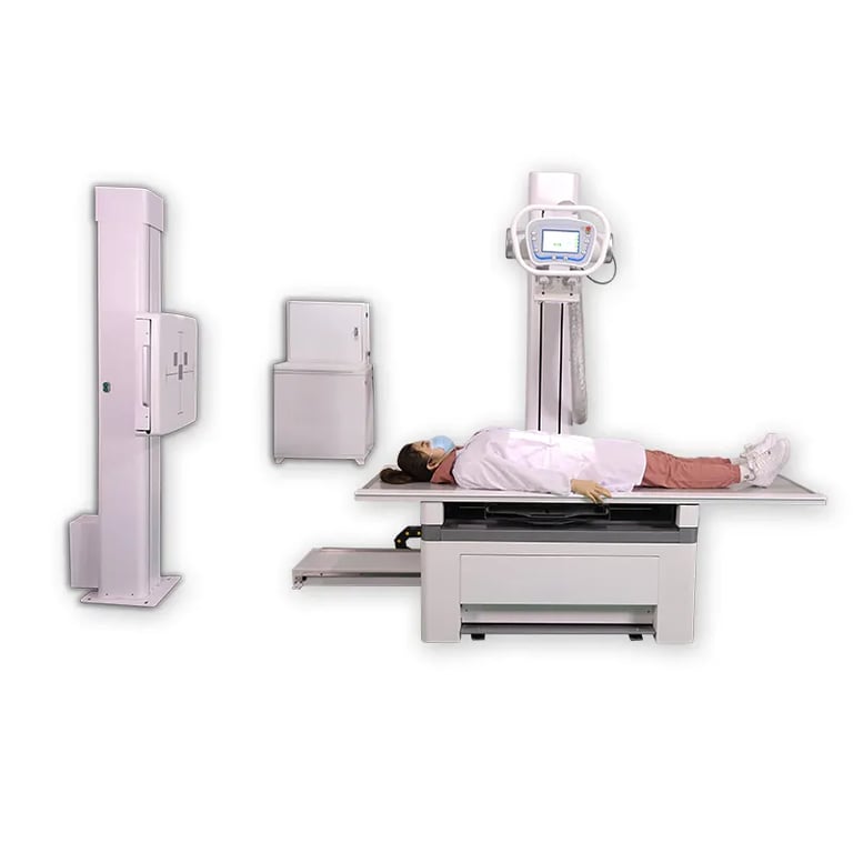
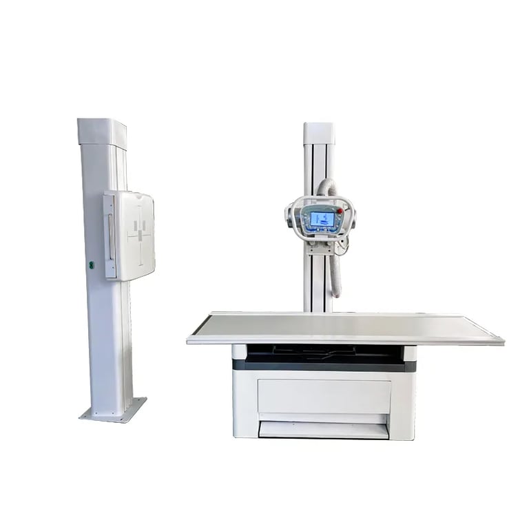
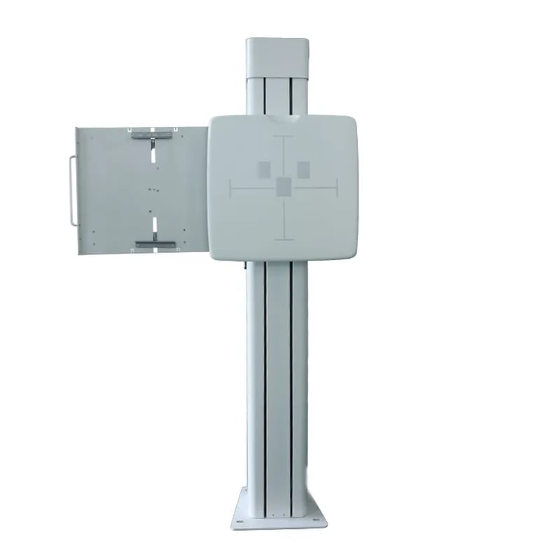
medical x ray machine orthopedics clinic 300mA digital x ray machine
A 300mA Digital X-ray Machine for orthopedic clinics is a powerful imaging device specifically designed to capture high-resolution X-ray images of bones and joints, essential for diagnosing musculoskeletal conditions. The 300mA (milliampere) designation indicates the machine’s ability to deliver a higher dose of X-rays, allowing for better image quality in orthopedic imaging, especially for large or dense areas of the body. These machines utilize digital radiography (DR) technology, which provides immediate, high-quality images with significantly reduced radiation exposure compared to traditional film-based systems.
This type of X-ray machine is widely used in orthopedic clinics, hospitals, and emergency care centers to assess bone fractures, joint problems, degenerative conditions, and more, aiding in both diagnosis and treatment planning. The digital nature ensures that images are instantly available for review, improving patient care and clinic workflow.


Key Features:
300mA X-ray Power:
The 300mA output provides sufficient power for capturing clear, detailed images of both dense and large anatomical areas, including the pelvis, spine, and large limb bones. This is especially important in orthopedic diagnostics where high-quality images of bones and joints are crucial.
Digital Radiography (DR) Technology:
Digital X-rays are processed and displayed almost instantly, eliminating the need for film development and providing immediate access to high-resolution images on a computer screen.
No chemical processing required, which is more environmentally friendly and reduces costs over time.
High-Resolution Imaging:
Digital detectors in 300mA X-ray machines provide superior image clarity, allowing for precise visualization of fractures, joint misalignments, arthritis, bone infections, and other orthopedic conditions.
Adjustable resolution settings ensure that both detailed images of small bones (e.g., fingers) and large structures (e.g., spine or hips) can be captured.
Low Radiation Exposure:
Digital systems use significantly less radiation compared to traditional X-ray machines, which is safer for both patients and healthcare providers. This is particularly important in an orthopedic setting where patients may need multiple imaging sessions.
Fast and Efficient Imaging:
Digital systems provide rapid imaging with immediate results, which reduces patient waiting time and streamlines the clinic's workflow.
The imaging process can be completed in a fraction of the time it takes with traditional film-based systems.
Enhanced Image Processing:
Advanced digital radiography systems include built-in image processing software that enhances image quality by adjusting contrast, brightness, and sharpness, improving diagnostic accuracy.
Features like zoom, rotation, and measuring tools are also available to examine images in greater detail.
Ergonomic Design:
Many modern digital X-ray machines have ergonomic designs with adjustable arms, movable tables, and intuitive touchscreens, making them easier for clinicians to use in an orthopedic setting.
Wireless or Networked Capabilities:
Many 300mA digital X-ray systems can connect to hospital or clinic networks, allowing easy sharing of images between departments, facilitating remote consultation, and improving collaboration.
Some models also have wireless detectors, providing flexibility and reducing the need for cables.
Long-Term Durability:
High-quality components are designed for heavy-duty usage, ensuring the machine can withstand the demands of an orthopedic practice with frequent imaging of patients.


Applications:
Bone Fractures:
The primary use of digital X-ray machines in orthopedic clinics is for diagnosing fractures. The high power (300mA) and resolution allow for clear imaging of fractures in both large bones (e.g., femur) and smaller bones (e.g., fingers and toes).
Joint Disorders:
Orthopedic X-rays are essential for assessing joint conditions, such as arthritis, joint deformities, dislocations, or conditions like osteoarthritis or rheumatoid arthritis that affect joint health over time.
Spinal Issues:
Spinal X-rays help diagnose conditions such as scoliosis, fractures, spinal degeneration, and herniated discs. The high-resolution images are crucial for detecting subtle abnormalities in the spine.
Bone Infections:
Digital X-ray is used to identify signs of infection in bones (osteomyelitis) and other bone-related pathologies, which is critical for early intervention and treatment.
Pre- and Post-Surgical Assessment:
For patients undergoing orthopedic surgery, X-rays are used to assess the alignment of bones and joints before surgery and to monitor healing after surgical procedures (e.g., after hip replacements or fracture fixation).
Sports Medicine:
Athletes commonly suffer from bone, joint, and ligament injuries, and orthopedic X-rays are essential for diagnosing conditions like stress fractures, ligament tears, and other trauma-related injuries.
Pediatric Orthopedics:
For pediatric patients, digital X-rays are used to assess bone growth and development, as well as to diagnose pediatric fractures, skeletal deformities, or developmental conditions like hip dysplasia.
Degenerative Diseases:
Conditions like osteoporosis and degenerative disk disease in older adults are diagnosed using digital X-ray imaging, helping to determine the severity of the condition and guide treatment options.


Why Choose a 300mA Digital X-ray Machine for Orthopedic Clinics?
Superior Image Quality:
The 300mA power level ensures that even large or dense bones and joints produce clear, detailed images, enabling orthopedic professionals to make accurate diagnoses and treatment decisions.
Quick Turnaround and Immediate Access:
With digital technology, the X-ray results are available almost immediately, allowing for quicker treatment decisions and reducing patient waiting times.
Reduced Radiation Exposure:
Digital radiography requires lower radiation doses compared to traditional film X-rays, making it safer for patients and medical staff, especially important in an orthopedic clinic where multiple X-rays might be required.
Cost Savings:
While the initial investment in digital X-ray equipment may be higher, it leads to long-term savings by eliminating the need for film, chemicals, and film processing equipment. Additionally, faster imaging leads to improved clinic efficiency and patient throughput.
Versatility and Efficiency:
The 300mA digital X-ray machine is versatile enough for a wide range of orthopedic imaging needs—from fractures to degenerative diseases to joint evaluations. The ease of use and fast image processing improve clinic workflows, making it suitable for busy practices.
Patient Comfort:
The quick and non-invasive nature of digital X-rays enhances patient comfort, as they do not need to wait for film development or endure long imaging sessions.
Advanced Imaging Software:
The integration of advanced software with digital X-ray systems allows for enhanced image processing, precise measurements, and manipulation (e.g., zoom, rotation), which is essential for accurate diagnostics in orthopedic care.
Long-Term Durability and Low Maintenance:
Digital X-ray machines are designed for longevity and require less maintenance than traditional film-based systems, which is crucial for maintaining consistent operation in a high-demand orthopedic clinic.
Ease of Integration with Other Systems:
These machines can be easily integrated with the clinic’s electronic medical records (EMR) or picture archiving and communication systems (PACS), streamlining patient management and improving workflow.


