
How to Read an X-Ray Image: A Basic Guide for Beginners
X-ray imaging is one of the most widely used diagnostic tools in medicine. It helps healthcare professionals to look inside the body, identify injuries, and diagnose various health conditions. But understanding how to read an X-ray image can seem daunting for those who are new to it. If you're a student in the medical field, a healthcare worker, or even a curious patient, this guide will help you break down the essential steps for reading an X-ray image. While mastering radiology requires years of training and experience, you can start by learning the basics of how to approach and interpret X-ray images.
2/21/20254 min read
1. Understand the Basics of X-Ray Technology
Before diving into interpreting X-ray images, it’s important to understand how the process works. When you get an X-ray, a machine sends a controlled amount of radiation through your body. The radiation is absorbed by different tissues in varying amounts, and the remaining radiation is captured by a detector or film on the other side of your body.
Dense materials, such as bones, absorb more radiation and appear white on the X-ray.
Less dense materials, like soft tissues, absorb less radiation and appear darker (black or gray).
The result is a two-dimensional image that displays the internal structure of your body, making it easier to identify fractures, infections, tumors, and other abnormalities.
2. Know the Basic Structure of an X-Ray Image
An X-ray image consists of several key components that you should understand:
Bone Structure: This is typically the easiest part of an X-ray to identify, as bones are dense and show up as white or light gray areas. Look for fractures, fractures, bone density issues (like osteoporosis), or other signs of bone damage.
Soft Tissues: These include muscles, fat, organs, and blood vessels. Soft tissues appear darker on an X-ray image compared to bones. Keep in mind that the details of soft tissues are usually not as clear on a standard X-ray, which is why additional imaging techniques like CT scans or MRIs are sometimes required.
Air: Air-filled spaces, like the lungs, appear black on an X-ray due to the minimal absorption of radiation. Airway obstructions or conditions like pneumonia can show up as lighter areas in the lungs.
Contrast Media: Sometimes, contrast agents are used in X-rays (like in a barium swallow or CT scan). These agents help highlight specific areas of interest, such as the digestive tract, and will appear bright white in the image.
3. Step-by-Step Process of Reading an X-Ray
Now, let’s break down the process of interpreting an X-ray step by step:
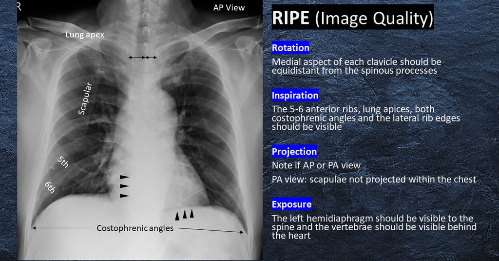
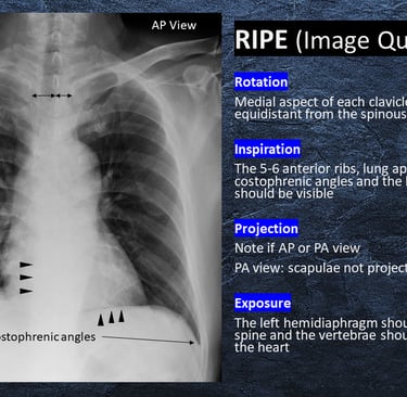
a. Start by Examining the Quality of the Image
Before diving into specifics, you need to evaluate the quality of the X-ray image. An unclear or poorly exposed X-ray can make it difficult to see the details necessary for accurate interpretation. Check for:
Proper exposure: Are all areas of interest clear and visible?
Good positioning: Is the patient’s body positioned correctly for the X-ray?
No motion blur: Is the image sharp, or does it look fuzzy due to patient movement?
b. Examine the Bones
Start with the bones, as they are the most straightforward to analyze. For example, in a chest X-ray, look at the rib cage, clavicles, spine, and shoulder bones. In an X-ray of a limb, check for:
Fractures: Look for breaks or cracks in the bone.
Deformities: Check for any abnormal bone alignment or shape.
Bone Density: Notice any areas that may appear lighter (indicating a decrease in bone density, like in osteoporosis) or darker (indicating a potential bone disease or tumor).
c. Look at the Soft Tissues
Next, focus on the soft tissues, keeping in mind that their appearance may be less detailed than bones. For example, in an abdominal X-ray:
Organs: Identify organs like the liver, spleen, and kidneys. Look for any enlargement, displacement, or unusual shapes.
Fluid or Air Collections: In cases like pneumonia or gastrointestinal obstructions, you might see abnormal fluid or air patterns in soft tissues.
Infections or Tumors: Look for any unusual dark or light areas that might indicate infection or masses.
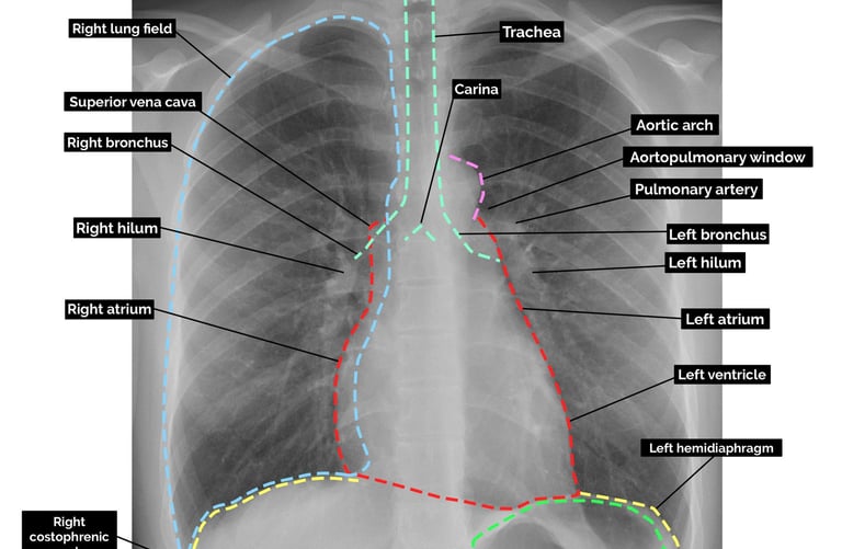
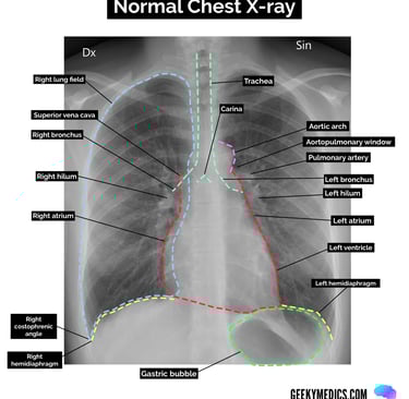
d. Evaluate the Lungs and Heart (in Chest X-Rays)
A chest X-ray typically includes the lungs and heart. Here’s what to look for:
Lungs: Check the lungs for any abnormal spots, shadows, or consolidations (which might indicate infection like pneumonia or other lung conditions).
Heart: Ensure that the heart’s size and shape appear normal. Enlargement or abnormalities could indicate conditions such as heart failure or pericardial disease.
e. Consider the Positioning and Symmetry
Always keep in mind the positioning of the X-ray. Is the body aligned properly? In a PA (posteroanterior) chest X-ray, for instance, you would want the patient’s chest to be in an upright position to avoid distortion. Compare both sides of the body, especially if it’s a full-body X-ray, to identify any asymmetry or abnormalities.
4. Look for Abnormalities
Once you've gone through the standard structure, now it’s time to focus on potential abnormalities. Some common signs to look out for include:
Fractures: Any bone breaks or cracks in the image.
Swelling or Inflammation: This can appear as areas of increased density or opacity.
Foreign Objects: Look for anything that doesn’t belong, such as bullets, shrapnel, or surgical hardware.
Abnormal Growths: Tumors or cysts will often show up as distinct, dark or light masses.
Diseases: Conditions like pneumonia, tuberculosis, or heart failure might reveal as abnormal shadows or fluid.
5. Consult a Radiologist
While the above steps can help you get started, interpreting X-rays accurately requires expertise. Always remember that reading X-ray images is a skill that comes with experience. Radiologists are specially trained to interpret these images, and their expertise ensures that diagnoses are accurate. If you notice something unusual or are unsure about an image, always consult a professional.
6. Practice Makes Perfect
The more you practice reading X-ray images, the better you’ll become. Whether you're a medical student, healthcare worker, or someone with an interest in radiology, hands-on practice, and continuous learning are key to improving your skills.


Reference Website Link:
RadiologyInfo.org – How to Read Your Chest X-ray Report
https://www.radiologyinfo.org/en/info/article-chest-xray-report?utm_source=chatgpt.com
The Radiology Assistant – Chest X-Ray Basic Interpretation
https://radiologyassistant.nl/chest/chest-x-ray/basic-interpretation?utm_source=chatgpt.com
TeachMeAnatomy – Plain Film X-Ray Principles and Interpretation
https://teachmeanatomy.info/the-basics/imaging/x-ray/?utm_source=chatgpt.com
RAI – Patient's Guide to Looking at an X-ray
https://4rai.com/2019/06/30/patients-guide-to-looking-at-an-x-ray/?utm_source=chatgpt.com
Mayo Clinic – X-ray: Imaging Test Quickly Helps Find Diagnosis
https://www.mayoclinic.org/tests-procedures/x-ray/about/pac-20395303?utm_source=chatgpt.com