How 3D X-Ray Imaging is Revolutionizing Diagnostic Accuracy
The world of medical imaging has undergone significant advancements over the past few decades, with one of the most transformative being the development of 3D X-ray imaging. This cutting-edge technology is reshaping the way healthcare professionals diagnose and treat various conditions by offering far greater clarity, precision, and depth than traditional 2D X-rays. As a result, 3D X-ray imaging is not only improving diagnostic accuracy but also enhancing patient outcomes and providing new possibilities for treatment planning. In this blog, we’ll explore how 3D X-ray imaging works, its benefits, and the areas where it is revolutionizing healthcare.
3/8/20254 min read


What is 3D X-Ray Imaging?
Traditional X-ray imaging produces a flat, two-dimensional (2D) image of the body. While this has been immensely useful for diagnosing a wide variety of conditions, it has its limitations. For example, overlapping structures can make it difficult to identify smaller or hidden abnormalities.
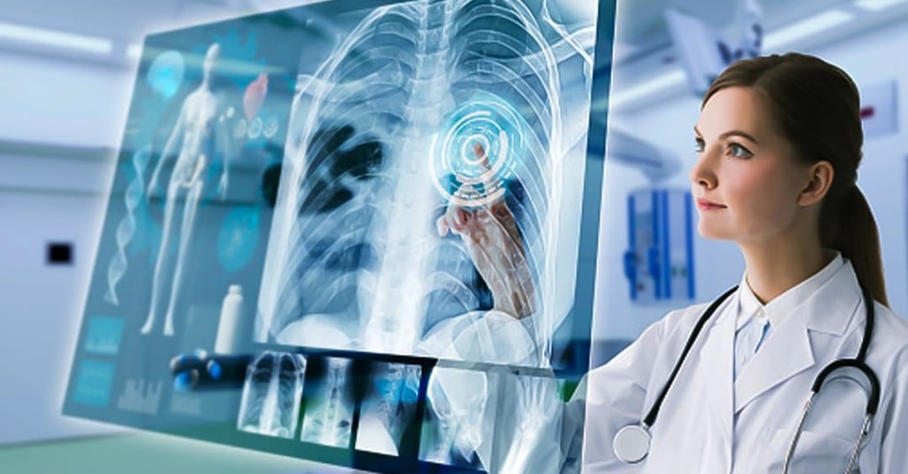
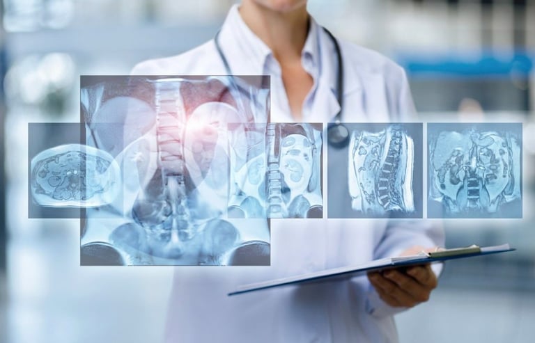
In contrast, 3D X-ray imaging (also known as tomosynthesis) captures multiple X-ray images from different angles, which are then processed by a computer to create a detailed, three-dimensional representation of the body’s internal structures. This allows healthcare professionals to view and analyze complex areas of the body from various angles, providing much clearer and more accurate information.
Key Benefits of 3D X-Ray Imaging
Enhanced Diagnostic Accuracy
One of the most significant advantages of 3D X-ray imaging is the improvement in diagnostic accuracy. With traditional 2D X-rays, overlapping tissues and structures can obscure critical details. However, with 3D imaging, doctors can examine the same area from multiple angles, significantly reducing the chance of misdiagnosis or missed abnormalities. For example, small tumors or fractures that might not be visible in a traditional 2D image can be clearly identified in 3D, leading to earlier detection and better outcomes.
Better Detection of Subtle Abnormalities
3D X-ray imaging is particularly valuable for detecting subtle abnormalities that might otherwise go unnoticed. Conditions such as small cancers, fractures, or bone deformities often require a level of detail that 2D X-rays cannot provide. By reconstructing a 3D image, healthcare professionals can spot these abnormalities more easily, making it possible to intervene at an earlier stage and significantly improving treatment prospects.
Minimized Risk of False Positives and False Negatives
3D imaging helps reduce the incidence of both false positives and false negatives, which are common issues with traditional 2D X-rays. A false positive occurs when a test result indicates the presence of an abnormality that isn’t there, leading to unnecessary procedures. A false negative, on the other hand, happens when a serious condition goes undetected because it wasn’t visible in a 2D image. With 3D X-ray imaging, the risk of both is greatly diminished, offering more reliable results.
Improved Treatment Planning and Precision
For complex medical cases, such as cancer, orthopedic conditions, or dental procedures, 3D imaging provides crucial insights that can help doctors plan treatments with greater precision. In oncology, for example, doctors can use 3D imaging to precisely locate and measure tumors, allowing for better-targeted treatment options. Similarly, for orthopedic surgeries, 3D X-ray imaging can help surgeons plan bone or joint surgeries with a more accurate understanding of the patient’s anatomy.
Reduced Radiation Exposure
Although 3D X-ray imaging uses multiple X-ray images, the overall radiation dose is often lower than that of traditional CT scans, which also produce detailed 3D images. This makes 3D X-ray imaging a safer option for patients, especially those who require frequent imaging, such as those with chronic conditions or those undergoing regular cancer screenings.
Applications of 3D X-Ray Imaging in Healthcare
The impact of 3D X-ray imaging spans across various medical fields. Here are some key areas where 3D X-ray is making a difference:
Breast Cancer Detection (3D Mammography or Tomosynthesis)
3D mammography, also known as digital breast tomosynthesis (DBT), has become a game-changer in breast cancer screening. By capturing multiple X-ray images from different angles, 3D mammography provides a more detailed and accurate view of the breast tissue. This improves the detection of tumors, particularly in women with dense breast tissue, where traditional 2D mammography might miss signs of cancer. 3D mammography also reduces the number of false positives, which means fewer unnecessary biopsies and anxiety for patients.
Orthopedics and Musculoskeletal Imaging
In orthopedics, 3D X-ray imaging allows for the detailed visualization of bones, joints, and soft tissues. It is especially useful in evaluating fractures, bone deformities, joint alignment issues, and conditions like osteoporosis. Surgeons can use 3D imaging to gain a better understanding of complex bone fractures and plan more accurate surgeries, leading to improved outcomes and faster recovery times.
Dental Imaging (3D Cone Beam CT)
In dentistry, cone beam computed tomography (CBCT) provides a 3D view of the teeth, jaws, and surrounding structures. CBCT is used for a variety of dental procedures, including implant planning, root canal treatments, and orthodontics. By offering a more detailed view than traditional 2D X-rays, CBCT helps dentists and oral surgeons make more accurate diagnoses and treatment plans.
Cardiology and Vascular Imaging
In cardiology, 3D X-ray imaging plays a critical role in assessing blood vessels, coronary arteries, and heart conditions. It’s particularly useful for planning and guiding interventions like angioplasty and stent placements. The ability to visualize the vasculature in 3D helps cardiologists make more accurate decisions during procedures, improving patient safety and reducing complications.
The Future of 3D X-Ray Imaging
As technology continues to evolve, 3D X-ray imaging is expected to become even more advanced. The integration of artificial intelligence (AI) and machine learning will further enhance the capabilities of 3D X-rays, enabling systems to automatically detect and analyze abnormalities with greater speed and precision. Additionally, with the development of portable 3D X-ray machines, healthcare providers can bring advanced diagnostic imaging to a wider range of settings, from hospitals to remote locations.
As the technology becomes more accessible and affordable, we can expect even more healthcare facilities to adopt 3D X-ray imaging, ultimately improving diagnostic capabilities and patient care worldwide.


American College of Radiology (ACR)
GE Healthcare
