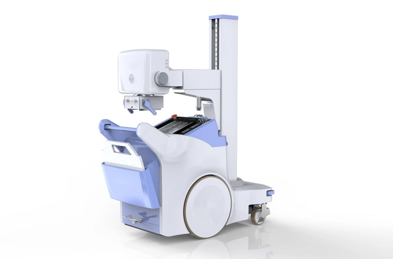
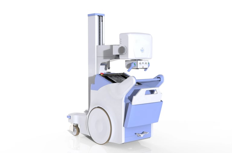
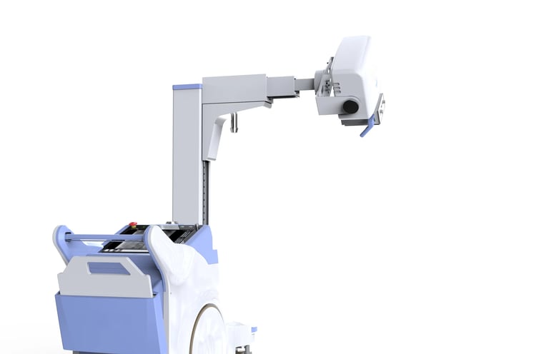
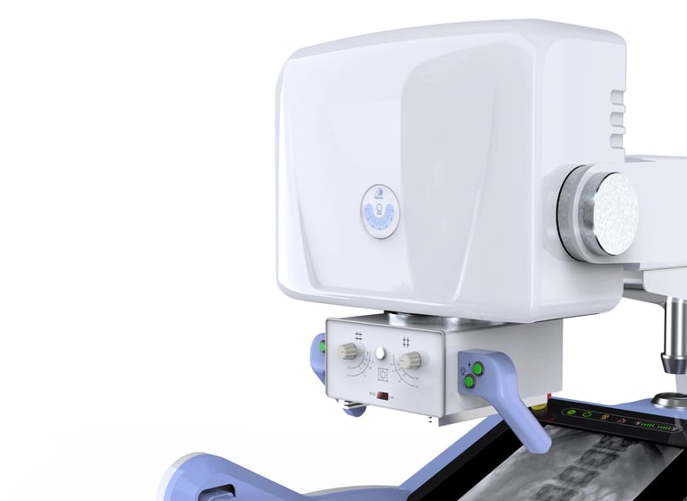
CE Digital mobile 500ma X-Ray DR system machine
The CE Digital Mobile 500mA X-ray DR System is a portable diagnostic imaging system designed to offer high-quality digital X-ray imaging with the flexibility and convenience of mobility. This X-ray system is CE certified, ensuring it meets European health, safety, and environmental protection standards. The 500mA output signifies its capability to generate powerful X-rays, making it suitable for a wide range of imaging applications, especially where high-resolution images are required.
This mobile system integrates digital radiography (DR) technology, enabling faster imaging, reduced radiation exposure, and immediate access to images. It is ideal for hospitals, clinics, emergency departments, where mobility and flexibility are critical for patient care.


Key Features:
500mA High-Power Output:
The 500mA output allows the machine to produce powerful X-rays, ensuring effective imaging of dense body parts such as the chest, abdomen, and bones.
Ideal for patients requiring deeper tissue penetration, including those with larger body types or complex diagnostic needs.
Digital Radiography (DR) System:
The machine is equipped with a digital radiography (DR) system, which captures high-quality images and displays them immediately on a monitor.
The digital system eliminates the need for traditional film, saving time and resources by reducing the processing time and the need for chemical agents.
Mobile and Compact Design:
The mobile design allows for flexibility in usage. The system can be easily moved to different areas within a hospital, clinic, or emergency room, making it ideal for bedside imaging or imaging of patients who cannot easily move.
Its compact nature makes it easier to store and transport, even in facilities with limited space.
Instant Image Acquisition:
The digital system provides real-time results, allowing healthcare providers to view images on a screen almost immediately after the exposure.
The image quality can be enhanced using digital tools like contrast adjustment, zoom, and sharpening, ensuring accurate diagnostics.
Low Radiation Exposure:
The system uses Automatic Exposure Control (AEC) to adjust the exposure parameters based on the patient's size and the body part being imaged. This ensures the lowest possible radiation dose while still providing clear, diagnostic images.
Digital radiography reduces radiation exposure compared to traditional X-ray methods, which is especially important for repeated imaging in sensitive populations (e.g., pediatric, geriatric patients).
Wireless Communication:
Many digital mobile systems allow for wireless transfer of images to the PACS (Picture Archiving and Communication System) or other storage platforms. This facilitates easy access and sharing of images among healthcare providers.
Advanced Image Processing:
The system often includes software with advanced image processing capabilities, allowing for adjustments in contrast, brightness, and sharpness, enhancing diagnostic capabilities for a variety of clinical conditions.
Image storage and retrieval are simplified with digital storage solutions, improving workflow efficiency in busy healthcare environments.
User-Friendly Interface:
The system typically features a touchscreen or intuitive control panel, making it easy for radiologists and technicians to operate the system without complicated settings.
Preset protocols for different imaging tasks can help ensure the correct exposure settings for specific areas of the body.
Flexible Imaging:
The mobile X-ray system is suitable for a range of imaging positions and body parts, whether lateral, frontal, or oblique views. It’s ideal for capturing a comprehensive set of images in one session, reducing patient handling time.


Applications:
Orthopedic Imaging:
Commonly used in diagnosing bone fractures, joint dislocations, and degenerative bone diseases such as arthritis.
It is ideal for trauma care, allowing for quick imaging of fractures or joint injuries in patients who may have limited mobility.
Chest Imaging:
The 500mA system is well-suited for chest X-rays to assess conditions such as pneumonia, lung infections, heart enlargement, and pulmonary diseases like emphysema or tuberculosis.
Used in cardiopulmonary imaging to detect heart or lung-related conditions.
Abdominal Imaging:
The system is effective in imaging the abdominal cavity, helping to detect conditions such as intestinal obstructions, gastrointestinal disorders, tumors, or organ anomalies.
It is useful for diagnosing kidney stones, liver conditions, or bladder problems.
Trauma and Emergency Imaging:
The portability and immediate image capture make it ideal for emergency rooms and trauma care.
Whether in field settings, ambulances, or emergency departments, this system can be used to quickly assess fractures, internal injuries, and head trauma.
Pediatric Imaging:
The low radiation dose makes the mobile 500mA X-ray machine suitable for pediatric imaging, which requires sensitive imaging techniques due to the vulnerability of children to radiation exposure.
It can be used for imaging bone fractures, pulmonary conditions, and abdominal disorders in children.
Hospital Bedside Imaging:
The mobile design allows for bedside imaging, which is particularly useful for patients who are immobile or critically ill. It eliminates the need to transport patients to radiology departments, thus reducing stress and complications.


Why Choose a CE Digital Mobile 500mA X-ray DR System?
Portability and Flexibility:
Its mobile design makes it highly versatile, allowing the system to be moved to different areas within a medical facility or even used in field-based applications (e.g., ambulances, rural areas, or emergency situations).
This mobility is ideal for hospitals and clinics with limited space or in scenarios where patients cannot easily be transported.
High-Quality Imaging:
The 500mA output ensures that this machine can produce high-resolution images, essential for accurate diagnosis, whether for orthopedic, pulmonary, or abdominal imaging.
The integration with digital radiography (DR) allows for real-time images with enhanced clarity and precision.
Lower Radiation Exposure:
The Automatic Exposure Control (AEC) ensures optimal image quality while minimizing radiation exposure, making it safer for both patients and healthcare workers.
Digital radiography also reduces overall radiation levels compared to traditional film-based X-rays.
Faster Diagnosis and Treatment:
The immediate availability of high-quality images helps healthcare professionals make faster decisions, particularly in emergency or trauma cases.
The system also enables rapid adjustments to imaging protocols, which can be critical in emergency situations.
Cost-Effective:
While the initial cost of the system may be higher, the digital radiography capabilities save on film, processing chemicals, and maintenance costs, reducing long-term operational costs.
Its portability also allows for multi-location use, maximizing the investment in the equipment.
Ease of Use:
The user-friendly interface and simple controls make this system easy to operate, reducing the learning curve for new users and increasing operational efficiency.
Versatility:
This system is ideal for a wide variety of medical imaging needs, from trauma care to routine checkups, pediatric imaging, and chest X-rays.
Its ability to perform multiple imaging views (lateral, oblique, and frontal) makes it a flexible solution for many types of procedures.
Reliable and Durable:
Designed to meet CE safety standards, this system is built to last and perform reliably in busy healthcare environments.


