
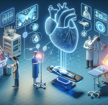
AI in X-Ray Imaging: Revolutionizing Diagnosis and Patient Care
Artificial Intelligence (AI) is dramatically improving the effectiveness of medical imaging, including X-rays. Traditionally, X-ray imaging has been one of the most widely used diagnostic tools in medicine, enabling healthcare professionals to detect fractures, infections, tumors, and other conditions. However, the interpretation of X-ray images can be complex and time-consuming, often requiring significant expertise from radiologists. This is where AI comes in, enhancing diagnostic accuracy, reducing human error, and speeding up the process. In this blog, we’ll explore how AI is transforming X-ray imaging and its potential to revolutionize healthcare.
4/5/20254 min read
1. AI in X-ray Image Interpretation: Enhancing Accuracy and Speed
The most significant application of AI in X-ray imaging is the interpretation of X-ray images. AI algorithms, particularly deep learning models, can analyze X-ray images at a much faster rate than human radiologists. These systems are trained using vast datasets of medical images, learning to identify patterns and abnormalities that are often invisible to the human eye.
Detecting Abnormalities and Pathologies: AI can be used to detect a variety of conditions in X-ray images, such as fractures, pneumonia, tuberculosis, and tumors. For example, AI systems can identify signs of lung cancer, breast cancer (in mammograms), and osteoporosis with remarkable precision. Studies have shown that AI models can sometimes match or even outperform human experts in detecting certain conditions, particularly when analyzing complex or subtle features in X-rays.
Consistency in Interpretation: One of the key advantages of AI in X-ray imaging is the ability to provide consistent and reproducible results. Human radiologists may occasionally miss small or subtle abnormalities, especially under high workloads or with less clear images. AI systems, on the other hand, are not subject to fatigue and can offer consistent interpretation, which can reduce the risk of missed diagnoses.
Speed and Efficiency: AI can analyze X-ray images in real-time, providing immediate results that assist in faster decision-making. This is especially important in emergency situations, where rapid diagnosis is critical. For instance, AI-powered X-ray systems can quickly identify pneumonia or fractures, allowing doctors to start treatment almost immediately, even before a radiologist’s review.
2. AI-Assisted X-ray for Early Disease Detection
AI is not just improving the interpretation of X-ray images; it is also enhancing the ability to detect diseases in their early stages. Early detection is crucial for effective treatment, and AI is proving to be a valuable tool in identifying diseases long before they become symptomatic.
Early Detection of Lung Cancer: In the case of lung cancer, AI has demonstrated the ability to identify small, early-stage tumors that may not be visible to the human eye on a traditional X-ray. For example, AI systems developed by companies like Google Health have shown promise in detecting lung cancer at a much earlier stage than conventional methods, which can significantly improve patient outcomes.
Tuberculosis (TB) Screening: AI-powered X-ray systems are being deployed to help detect tuberculosis (TB) in areas where access to healthcare is limited. In regions where TB is prevalent, AI can be used to analyze chest X-rays and identify signs of the disease, even in its early stages. This early detection can lead to more timely treatment and a reduction in transmission rates.
Breast Cancer Detection (Mammography): AI is also being integrated into mammography, an essential X-ray imaging technique for breast cancer screening. AI algorithms can detect subtle abnormalities in mammograms, improving early detection rates and reducing false positives or false negatives.
3. AI in X-ray Quality Control: Ensuring Optimal Imaging
While AI is often used to analyze the content of X-ray images, it can also be employed to ensure that the images themselves are of the highest quality. High-quality images are essential for accurate diagnoses, and AI systems are capable of optimizing image quality in several ways.
Automatic Image Enhancement: AI can help enhance the quality of X-ray images by adjusting factors like contrast, brightness, and resolution. This is especially useful when images are of lower quality due to technical limitations, such as patient movement or insufficient exposure. AI systems can automatically detect poor-quality images and correct them, making it easier for radiologists to analyze.
Image Denoising: AI algorithms can also be used to remove noise from X-ray images, improving the clarity and accuracy of the final result. This is particularly useful in low-dose X-ray imaging, where the radiation exposure is intentionally reduced to minimize risk, but the images may appear grainy. AI can help enhance these low-dose images without compromising the diagnostic value.
Optimizing Exposure Levels: AI can monitor exposure levels during X-ray imaging to ensure that patients receive the optimal amount of radiation. Overexposure can be harmful, while underexposure can result in poor image quality. AI algorithms can help radiology technicians adjust settings to achieve the best balance between image quality and radiation exposure.
4. AI in X-ray Screening: Access and Scalability
AI’s role in X-ray imaging is also expanding beyond traditional hospital settings, making high-quality diagnostics more accessible in remote or underserved areas.
Telemedicine and Remote Screening: AI-powered X-ray interpretation tools are being integrated into telemedicine platforms, allowing healthcare professionals in remote areas to send X-ray images to AI systems for analysis. These platforms can then provide automated interpretations, allowing doctors in these regions to make informed decisions without needing an expert radiologist on-site. This is especially valuable in rural or resource-limited areas where access to skilled radiologists may be limited.
Mass Screening Programs: AI can also be used in large-scale screening programs for diseases such as lung cancer, tuberculosis, and breast cancer. By automating the analysis of thousands or even millions of X-ray images, AI systems can help healthcare organizations rapidly identify individuals who need further evaluation, improving early detection and saving time.
5. Future Potential of AI in X-ray Imaging
As AI technology continues to evolve, we can expect even more advanced applications in the field of X-ray imaging. Here are a few future developments we might see:
AI-Driven Radiology Assistants: AI could evolve into a “virtual assistant” for radiologists, providing not only automated analysis of X-ray images but also suggesting potential diagnoses and treatment pathways. This could allow radiologists to focus on more complex cases while ensuring that routine examinations are completed quickly and accurately.
Integration with Other Imaging Modalities: In the future, AI could combine data from X-rays, MRIs, CT scans, and other imaging technologies to provide a more comprehensive analysis of a patient’s condition. This integrated approach could improve diagnostic accuracy and lead to better patient care.
AI for Predictive Analytics: By analyzing large datasets of X-ray images alongside patient data, AI could predict the likelihood of certain conditions developing in the future. This would enable doctors to take proactive measures, such as preventative treatments or lifestyle changes, to avoid the onset of disease.
Reference Website Links:
Google Health and AI in Medical Imaging
Radiological Society of North America (RSNA)
AI in Healthcare by Mayo Clinic
Nature - AI in X-ray Imaging
Medical Imaging and AI - NVIDIA
Siemens Healthineers - AI in Imaging