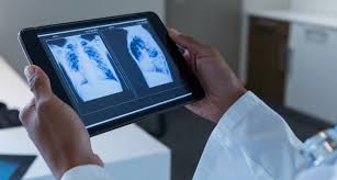
AI and Machine Learning in X-ray Imaging: A Revolution in Diagnostics
Artificial Intelligence (AI) and Machine Learning (ML) are making significant strides in the world of medical imaging, and X-ray technology is no exception. These cutting-edge technologies are improving diagnostic accuracy, enhancing workflow efficiency, and reducing the burden on healthcare professionals. Let’s take a closer look at how AI and ML are transforming X-ray imaging.
4/2/20253 min read


How AI and ML Are Being Integrated into X-ray Imaging
AI and ML algorithms are trained to recognize patterns in X-ray images that may be too subtle for the human eye to detect. By analyzing thousands (or even millions) of images, these systems learn to identify specific conditions such as fractures, infections, lung diseases, or even early-stage cancers with incredible precision.
Machine learning models can be trained using annotated datasets, where experts label images with the correct diagnoses. Over time, the algorithm "learns" from these labels and becomes capable of diagnosing new X-ray images with increasing accuracy.
Improved Diagnostics and Early Detection
One of the most compelling benefits of AI in X-ray imaging is its potential for early detection. In medical imaging, time is of the essence. Catching a disease or condition early significantly improves treatment outcomes, and AI can be a powerful tool in identifying these issues even in their earliest stages.
For example, AI algorithms are being used to detect conditions like pneumonia, fractures, and even lung cancer in chest X-rays. These algorithms can identify minor changes in tissues or structures that might otherwise be overlooked, offering doctors a more detailed perspective to guide their diagnostic decisions.
In a study published by Radiology (a leading medical journal), AI models were able to detect lung cancer in chest X-rays more effectively than radiologists in certain cases, showing just how powerful these tools can be in assisting healthcare professionals.
Streamlining Radiologist Workflows
X-ray imaging has traditionally been time-consuming, with radiologists needing to review hundreds or even thousands of images each day. This can lead to burnout and increased chances of missing key details due to fatigue.
AI helps alleviate this burden by providing automated analysis that assists radiologists in reviewing X-ray images faster. For example, AI tools can automatically highlight areas of concern in an X-ray, such as a tumor or a crack in the bone, making it easier for the radiologist to focus their attention on critical issues.
These automated systems also help prioritize cases based on urgency. A patient with a potential life-threatening condition, such as a massive lung infection, can be flagged for quicker review, ensuring that they receive timely care.
Reducing Human Error
One of the biggest challenges in medical diagnostics is human error. Even the most skilled radiologists can occasionally miss a subtle anomaly due to fatigue, distractions, or the sheer volume of images they need to review. AI and ML models, on the other hand, are not subject to the same physical limitations and can consistently provide accurate and reliable results.
By assisting radiologists and even providing a second opinion, AI can reduce diagnostic errors, leading to better outcomes for patients and more confidence for healthcare providers.
Real-World Applications
Several AI-powered X-ray tools are already in use today, and they are being integrated into clinical practices around the world. Here are some examples:
Zebra Medical Vision: This AI company has developed a tool that automatically analyzes medical imaging to detect a variety of conditions, including cardiovascular diseases, cancers, and fractures. Their software uses deep learning algorithms to identify abnormalities in X-rays and other imaging modalities.
Lunit INSIGHT: This is an AI-powered platform designed specifically for chest X-rays. It uses deep learning algorithms to detect lung diseases such as tuberculosis and pneumonia, as well as lung cancer. The platform has been deployed in various hospitals and healthcare settings to enhance diagnostic workflows.
Google Health’s AI for Mammography: Google Health has developed an AI system that can interpret mammograms for breast cancer detection. The AI system has been shown to outperform radiologists in certain situations, suggesting that AI can improve early detection rates in breast cancer diagnoses.
Challenges and Considerations
Despite the incredible promise AI and ML hold for X-ray imaging, there are still several challenges to address. One key issue is ensuring that the algorithms are generalizable—meaning they must work effectively across diverse patient populations, including those from different racial, ethnic, and demographic groups. AI models need to be trained on a wide range of data to avoid biases that could lead to incorrect diagnoses for certain groups of patients.
Additionally, the integration of AI in healthcare requires robust regulatory oversight to ensure that these tools meet medical safety standards. As AI continues to evolve, it is important for healthcare providers to maintain a balance between trusting automated systems and using their expertise to make final clinical decisions.
The Future of AI in X-ray Imaging
Looking ahead, AI is likely to play an even more prominent role in X-ray imaging. With improvements in algorithm accuracy, faster processing speeds, and greater integration with other healthcare technologies, AI-powered X-ray tools will continue to enhance diagnostic capabilities.
Moreover, as AI and machine learning models continue to learn and adapt from new data, their potential to detect complex medical conditions will only grow. This could open up new possibilities for personalized medicine, where treatment plans are tailored based on more precise and early diagnoses.
Reference Website Links:
Zebra Medical Vision
Lunit INSIGHT
Google Health’s AI for Mammography
Radiology Society of North America (RSNA)
Nature Medicine – AI in Medical Imaging
PubMed Central – AI in Medical Imaging
