300 Ma Hospital X-ray Digital X-ray Radiography X-Ray Machine
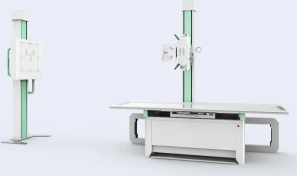
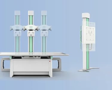
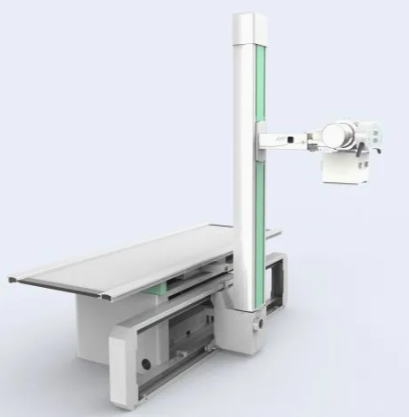
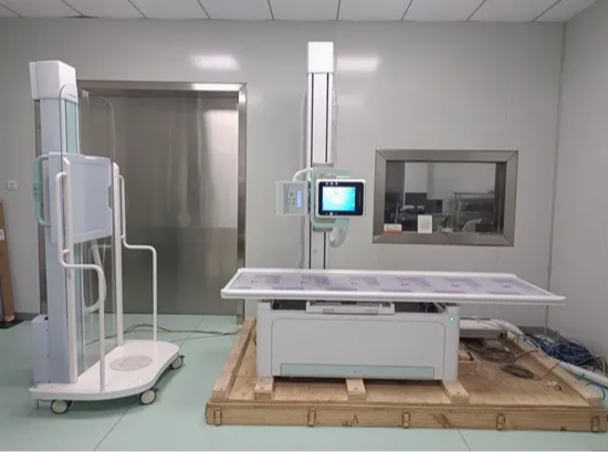
The 300mA Hospital X-ray Digital Radiography System is designed to deliver high-quality X-ray images, utilizing the latest digital radiography (DR) technology. The system integrates advanced digital sensors to provide sharp, clear, and immediate images, reducing the time required for diagnosis. This machine is used primarily in hospitals, diagnostic centers, and clinics for a variety of medical imaging procedures, including imaging of bones, joints, chest, and abdominal areas.


Key Features:
300mA Power Output: The machine features a high tube current of 300mA, allowing for high-resolution imaging of both dense and delicate tissues. This provides fast imaging results while maintaining excellent image quality, making it suitable for a broad range of diagnostic applications.
Digital Radiography (DR) System: Equipped with digital sensors, the system captures high-quality images instantly. Digital images are processed quickly, displayed on a monitor, and can be easily stored and transferred to electronic health record (EHR) systems for efficient diagnosis and treatment planning.
Low Radiation Exposure: The system is designed with optimized exposure settings to ensure minimal radiation exposure to patients while still delivering high-quality images. This aligns with modern radiation safety standards and helps reduce the risk of radiation-related issues.
Automatic Exposure Control (AEC): The system includes Automatic Exposure Control, which adjusts the exposure parameters based on the size and density of the area being imaged. This ensures optimal image quality for each patient while minimizing unnecessary radiation.
High Image Resolution: The digital imaging system provides superior contrast, clarity, and detail, which are essential for detecting fractures, tumors, infections, and other medical conditions. The high resolution allows for precise diagnostics.
Wide Imaging Range: The system is versatile and can be used for imaging various body parts, such as the chest, bones, joints, spine, abdominal organs, and extremities. It is capable of imaging both dense areas (e.g., bones) and softer tissues, such as the lungs or abdominal organs.
Fast Image Processing: Digital radiography allows for immediate viewing of images after exposure, reducing the time between taking an X-ray and the diagnosis. This is particularly useful in emergency situations or where quick decision-making is needed.
Computerized Image Storage and Retrieval: The digital X-ray images are stored electronically, making it easy to access and retrieve them for future reference. Images can be shared electronically with other healthcare providers for collaboration or second opinions.
Ergonomically Designed: The machine is designed for ease of use and comfortable operation by radiologists or technicians. The ergonomic design helps to improve workflow, reduce operator fatigue, and enhance overall productivity.
Space-Saving Design: Modern digital X-ray machines are often designed to be more compact than older analog systems, saving space in hospital rooms or imaging centers while still providing high-quality diagnostic imaging.


Applications:
Routine Medical Imaging: The system is commonly used for routine diagnostic imaging in hospitals and clinics. It is ideal for imaging bones (e.g., fractures), joints, and the chest to evaluate lung and heart conditions.
Emergency Medicine: In emergency departments, rapid and high-quality imaging is essential for diagnosing trauma, fractures, dislocations, or internal injuries. The 300mA digital X-ray machine provides clear, immediate images for faster diagnosis and treatment.
Orthopedic Imaging: This system is perfect for capturing detailed images of bones and joints, helping to diagnose fractures, arthritis, osteoporosis, bone infections, and other musculoskeletal disorders.
Chest Imaging: The system is often used for chest X-rays to diagnose lung conditions (e.g., pneumonia, tuberculosis, lung cancer), heart conditions (e.g., heart failure, enlarged heart), and other thoracic diseases.
Abdominal Imaging: This system is effective for imaging abdominal organs, such as the liver, kidneys, and intestines, to diagnose conditions like kidney stones, gastrointestinal problems, and tumors.
Spinal Imaging: The system is frequently used to obtain detailed images of the spine to diagnose conditions such as scoliosis, spinal fractures, herniated discs, or other spinal disorders.
Pre-Surgical Planning: Surgeons often use X-ray images to plan operations, such as orthopedic surgeries, spinal surgeries, and soft tissue procedures, to ensure the most accurate approach.
Cancer Screening and Monitoring: It is widely used in cancer diagnosis and monitoring, helping to detect and evaluate the size and spread of tumors, particularly in the lungs, bones, and abdominal regions.


Why Choose the 300mA Hospital X-ray Digital Radiography System?
High Image Quality: With 300mA output and advanced digital sensors, the system delivers high-resolution, clear, and detailed images, essential for accurate diagnoses, even in complex cases.
Reduced Radiation Exposure: The system is designed to minimize radiation exposure while still delivering optimal imaging results. This makes it safer for patients and healthcare providers, in compliance with current safety regulations.
Fast and Efficient Workflow: The digital nature of the system allows for rapid image capture and immediate processing. This reduces patient wait times and improves the speed of diagnosis, particularly in emergency settings.
Cost-Effective: While the initial investment may be higher than traditional film-based X-ray systems, the long-term savings from digital imaging (such as eliminating the need for film, chemicals, and storage space) make it a cost-effective choice for hospitals and clinics.
Ease of Use: The system’s user-friendly interface ensures that technicians can operate the equipment with minimal training. The image processing is straightforward, and the system includes features like automatic exposure control for consistent results.
Space Efficiency: The compact design of modern digital X-ray systems allows for installation in smaller spaces, which is ideal for hospitals with limited room for large equipment.
Electronic Image Storage: Digital radiography enables the storage of images in electronic formats, making it easy to share, store, and retrieve patient images. This enhances record-keeping, improves collaboration, and reduces the risk of losing physical images.
Remote Access and Sharing: The system’s digital images can be shared electronically with other healthcare providers, enabling quick consultations and second opinions, even remotely.
Durability and Reliability: Modern digital X-ray systems are built to be durable and reliable, offering long-term performance with minimal maintenance.


